Inicio Blog Página 42

New findings explain the connection between melatonin and type 2 diabetes

0

Melatonin is a naturally occurring hormone that helps maintain our circadian rhythm. The amount of melatonin varies throughout the course of the day, and is affected by light. When it’s dark, the level of melatonin increases, peaking at night. It is often referred to as “the hormone of darkness”, and used as a sleeping drug or to prevent jet lag, among other things.

“A third of all people carry this specific gene variant. Our results show that the effect of melatonin is stronger in them. We believe that this explains their increased risk of developing type 2 diabetes”, says Hindrik Mulder.

The findings, which are published in the scientific journal Cell Metabolism, are the result of many years of work. Already in 2009, the researchers behind this new study were able to present an extensive gene mapping study that showed that the gene variant of the melatonin receptor 1B, which is common in the population, increases the risk of type 2 diabetes. The gene variant causes the level of the melatonin receptor on the insulin cell surface to increase, which makes the cells become more sensitive to melatonin and impairs their ability to secrete insulin.

The researchers have now moved on to study the processes in mice and human beta cells, as well as completed a study of how the effects of drugs are influenced by genetic factors – one of the first of its kind within type 2 diabetes, where participants have been recruited based on their genotype, that is, their genetic make-up.

The study included 23 healthy people and carriers of the gene variant in question and 22 non-carriers. All participants were roughly of the same age and with the same body mass index (BMI). There was also no difference in terms of their family history of diabetes.

They were given four milligrams of melatonin before bedtime over the course of three months.

Among other things, the study showed that:

  • insulin secretion was significantly lower among those who carried the risk gene than those in the control group.
  • the glucose (sugar) concentration in the blood was higher among all participants after being treated with melatonin for three months. However, it was especially evident in carriers of the risk gene who were unable to increase their insulin secretion.

It has been previously known that people who work overnight shifts suffer from metabolic diseases such as type 2 diabetes to a greater extent.

“It is perhaps therefore less suitable for carriers of the risk gene to work overnight shifts, as the level of melatonin will probably increase at the same time as the effects of the increase are enhanced. There is still no scientific support for this theory, but it ought to be studied in the future, on the basis of our new findings”, argues Hindrik Mulder.

Source: Lund University

Listado de emisiones anteriores

No se encontraron entradas.

New PSA test examines protein structures to detect prostate cancers

0

The study – part of an ongoing multicenter prospective clinical trial – found that the IsoPSATM test can also differentiate between high-risk and low-risk disease, as well as benign conditions.

Although widely used, the current PSA test relies on detection strategies that have poor specificity for cancer – just 25 percent of men who have a prostate biopsy due to an elevated PSA level actually have prostate cancer, according to the National Cancer Institute – and an inability to determine the aggressiveness of the disease.

The IsoPSA test, however, identifies prostate cancer in a new way. Developed by Cleveland Clinic, in collaboration with Cleveland Diagnostics, Inc., IsoPSA identifies the molecular structural changes in protein biomarkers. It is able to detect cancer by identifying these structural changes, as opposed to current tests that simply measure the protein’s concentration in a patient’s blood.

“While the PSA test has undoubtedly been one of the most successful biomarkers in history, its limitations are well known. Even currently available prostate cancer diagnostic tests rely on biomarkers that can be affected by physiological factors unrelated to cancer,” said Eric Klein, M.D., chair of Cleveland Clinic’s Glickman Urological & Kidney Institute. “These study results show that using structural changes in PSA protein to detect cancer is more effective and can help prevent unneeded biopsies in low-risk patients.”

The clinical trial involves six healthcare institutions and 132 patients, to date. It examined the ability of IsoPSA to distinguish patients with and without biopsy-confirmed evidence of cancer. It also evaluated the test’s precision in differentiating patients with high-grade (Gleason = 7) cancer from those with low-grade (Gleason = 6) disease and benign findings after standard ultrasound-guided biopsy of the prostate.

Substituting the IsoPSA structure-based composite index for the standard PSA resulted in improvement in diagnostic accuracy. Compared with serum PSA testing, IsoPSA performed better in both sensitivity and specificity.

“We took an ‘out of the box’ approach that has shown success in detecting prostate cancer but also has the potential to address other clinically important questions such as clinical surveillance of patients after treatment,” said Mark Stovsky, M.D., staff member, Cleveland Clinic Glickman Urological & Kidney Institute’s Department of Urology. Stovsky has a leadership position (Chief Medical Officer) and investment interest in Cleveland Diagnostics, Inc. “In general, the clinical utility of prostate cancer early detection and screening tests is often limited by the fact that biomarker concentrations may be affected by physiological processes unrelated to cancer, such as inflammation, as well as the relative lack of specificity of these biomarkers to the cancer phenotype. In contrast, clinical research data suggests that the IsoPSA assay can interrogate the entire PSA isoform distribution as a single stand-alone diagnostic tool which can reliably identify structural changes in the PSA protein that correlate with the presence or absence and aggressiveness of prostate cancer.”

Source: Sciencedaily.com

Study finds no change in antibody levels associated with food allergy

0

The new data, published ahead of print in the April 18 edition of Journal of Allergy and Clinical Immunology, suggest that the increases in prevalence of food allergies over the past several decades may be due to either an increase in the recognition and diagnosis of food allergy or to a changing relationship between the presence of IgE and food allergy symptoms.

“Since the 1990s, studies have shown the prevalence of food allergies in children has grown by at least 50 percent,” says senior study author Corinne A. Keet, M.D., Ph.D., associate professor of pediatrics at the Johns Hopkins University School of Medicine. “Today, we think about 5 percent of children in the Unites States have a food allergy, with the most common triggering allergens found in milk, eggs, shellfish and peanuts. Why allergies have seemed to increase, however, has been unclear and has led to a significant amount of research on causes underlying the food allergy ‘epidemic.'”

As the number of children with reported food allergies has risen, the authors wondered whether the number of children who have the IgE antibodies to food that are essential for food allergy to occur had risen as well. To answer this question, they analyzed data from nearly 8,000 children ages 6 through 19 as part of the National Health and Nutrition Examination Survey. Some samples were collected between 1988 and 1994 and recently analyzed for food-specific IgE by the authors, and others were collected between 2005 and 2006, with food-specific IgE measured at that time.

Overall, the team found no increase in the number of children sensitized to peanuts, milk or eggs, while the number with antibodies to shrimp actually decreased. “We were really very surprised,” says Keet. “It hasn’t only been self-reported food allergy rates that have risen but emergency room visits and hospitalizations for food allergies as well.

It’s important to know whether changing sensitization to foods underlies these trends, because if sensitization hasn’t changed, then continuing to look for environmental factors that affect sensitization may not answer why food allergy has increased.”

One possible explanation for the results, Keet says, is that parents, patients and physicians are simply more aware of food allergies today than they were a few decades ago. “In the past, there may have been more people who just didn’t eat a food because it made them feel sick, but they didn’t necessarily call that an allergy,” Keet says. “Today, we may be much more likely to suspect allergy when a child has a rash or other symptoms after eating a certain food.”

Alternatively, scientists know that although food-specific IgE antibodies are essential for the kind of food allergy that leads to anaphylaxis, many people with IgE antibodies to foods can eat those foods without problems. “These results raise the question of whether something has changed in the relationship between food-specific IgE and clinical food allergy over the past few decades,” says Emily McGowan, M.D., assistant professor of medicine at the Johns Hopkins University School of Medicine and the first author of this study. “For example, a recent study showed that introducing peanuts early in infancy prevented most peanut allergies but didn’t really lead to decreased rates of having IgE to peanuts. We don’t really understand all of the reasons why one person with IgE to a food will have serious reactions to the food, while another can eat it without problems. This is an area that needs more research.”

The investigators say studies on more recent blood samples and broader groups of people would help confirm the finding and offer more details on the link between food-specific IgE levels and food allergies.

Source: Hopkins Medicine


New model for active surveillance of prostate cancer tested

0
Christopher Kane, M.D., is chair of the Department of Urology at UC San Diego Health.

The evidence-based approach uses best practices to appropriately select and follow patients to avoid disease overtreatment. Results of the three-year study are now published online in the journal of Urology.

“Active surveillance is a strategy that is recommended by physician and quality organizations to avoid the overtreatment of slow-growing prostate cancer,” said Christopher Kane, MD, senior author and chair of the Department of Urology at UC San Diego Health. “Acceptance of this strategy by patients and urologists, however, has lagged for a number of reasons. What we have developed is a safe method to enhance acceptance and use of this disease management approach.”

Active surveillance (AS) is the practice of closely monitoring slow, indolent forms of prostate cancer with prostate-specific antigen (PSA) blood tests, digital rectal prostate exams and, potentially, biopsies. Kane added that AS recognizes that there is a large group of men with a form of low-grade prostate cancer whose long-term survival is not impacted by non-treatment.

“With this new model, we were able to increase rates of surveillance to benefit patients through use of provider education and a standardized report card,” said Franklin Gaylis, MD, Chief Scientific Officer, Genesis Healthcare and the study’s first author. “With this university and private-practice research collaboration, we were able monitor 190 patients undergoing active surveillance while evaluating the effectiveness of our own individual clinical practices.”

Urologists at Genesis Healthcare and UC San Diego Health jointly developed a reporting mechanism to improve the process of tracking patients with prostate cancer. The research team developed standardized selection criteria based on scientific literature for patients to be followed with AS according to tumor characteristics, including clinical cancer staging, Gleason (pathology grading) and PSA scores. In addition, comparative dashboards were developed to show individual physician AS adoption rates compared to their peers.

“What we found is that active surveillance increased from 43.75 percent to 82.6 percent among the very low-risk patients,” said Gaylis, a voluntary professor in the Division of Urology at UC San Diego School of Medicine. “Besides this approach, enhancing quality of care through established standardized processes and outcomes feedback, there may be a benefit from a cost-savings perspective. This model may be particularly helpful as the U.S. switches from a volume to value-based system of care for reimbursement requiring physicians to improve quality while at the same time reducing cost.”

Prostate cancer is the most prevalent solid organ malignancy among American men, accounting for almost 30 percent of new cancer diagnoses. The National Institutes of Health estimates approximately 220,800 new cases of prostate cancer will be identified in 2015.


Source: sciencedaily.com

HPV infection can be identified in self-collected vaginal swabs

0

But self sampling with vaginal swabs may provide materials that screen as accurately as the more labor-intensive approach using cervical samples obtained by clinicians. This finding is critical to developing same day screening and treatment, which is key to ensuring that women with precancerous lesions are treated in this largely unconnected (electronically) country, and in others like it. The research appeared online April 13, 2016 in the Journal of Clinical Microbiology, which is published by the American Society for Microbiology.

“This is the first time there’s been a direct comparison between self-collected vaginal swabs and clinician-collected cervical specimens using a screening device that can provide same day results,” said corresponding author Andrew Vallely, PhD, Associate Professor, Public Health Interventions Research Group, The Kirby Institute, University of New South Wales, Sydney, Australia.

Evaluating self-collected samples was a critical milestone towards meeting the ultimate goal of this research: to enable same-day screening and treatment. Same-day treatment is needed in high-burden, low-income countries such as Papua New Guinea because when patients leave the clinic, it often becomes impossible to find them again, should their results show that they need treatment.

“The majority of the country’s population live in rural communities, many of which are very isolated,” explained Vallely, who is also Professorial Research Fellow, Sexual and Reproductive Health, Papua New Guinea Institute of Medical Research, Goroka. A dearth of roads, and difficult terrain exacerbates the isolation, as do limited or absent mobile phones and internet connectivity, and postal services that are very poor and unreliable.

Self sampling, combined with use of a new, high speed, fully automated molecular assay for high risk HPV infection, called the Xpert HPV Test, makes it possible to screen and treat patients in a single day. Self-sampling alleviates pressure on highly skilled clinical staff who would otherwise only be able to screen women by conducting a time-consuming pelvic examination to collect specimens, said Vallely.

“By readily identifying women who have a high risk HPV infection, this clinic-based self-sampling strategy would allow health services in low-income settings such as Papua New Guinea to focus their efforts on those women who are most at risk of cervical pre-cancer and cancer,” said Vallely.

Once those at increased risk are identified by the Xpert HPV Test, the final step in screening is to paint the screened women’s cervixes with acetic acid — vinegar, said Vallely. The acetic acid causes the precancerous lesions, which are not visible to the naked eye, to stain white. The clinicians then ablate the lesions using cryotherapy — a relatively non-invasive procedure to obliterate the abnormal tissues that allows the women to leave for home the same day. (Women who have developed malignancies are referred to specialists.)

Papua New Guinea has a very high burden of disease, said Vallely. The rate of new cases is six to seven times higher than in Australia and New Zealand, and mortality is around 14 times higher, making HPV-associated cancers a leading cause of premature death.

Source: Sciencedaily.com


Mass. General-developed device may provide rapid diagnosis of bacterial infections

0
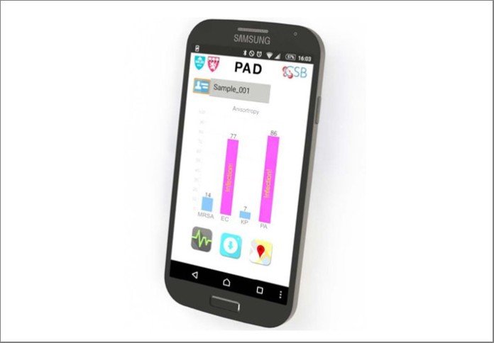
A smartphone readout from the Polarization Anisotropy Diagnostics (PAD) system reveals the pathogens responsible for an infection and factors such as antibiotic resistance (Chen-Han Huang, PhD, and Ki Soo Park, PhD, Center for System Biology, Massachusetts General Hospital)

A team of Massachusetts General Hospital (MGH) investigators has developed a device with the potential of shortening the time required to rapidly diagnose pathogens responsible for health-care-associated infections from a couple of days to a matter of hours. The system described in the journal Science Advances also would allow point-of-care diagnosis, as it does not require the facilities and expertise available only in hospital laboratories.

“Health-care-associated infections are a major problem that affects more than 600,000 patients each year, more than 10 percent of whom will die, and incurs more $100 billion in related costs,” says Ralph Weissleder, MD, PhD, director of the MGH Center for Systems Biology, Thrall Family Professor of Radiology at Harvard Medical School (HMS) and co-senior author of the report. “Rapid and efficient diagnosis of the pathogen is a critical first step in choosing the appropriate antibiotic regimen, and this system could provide that information in a physician’s office in less than two hours.”

While considered the gold standard for diagnosing bacterial infections, traditional culture-based diagnosis can take several days and requires specialized equipment, trained laboratory personnel and procedures that vary depending on the particular pathogen. Emerging genetic approaches that identify bacterial species by their nucleic acid sequences are powerful but still require complex equipment and workflows, restricting such testing to specialized hospital laboratories.

The system developed by the MGH team, dubbed PAD for Polarization Anisotropy Diagnostics, allows for accurate genetic testing in a simple device. Bacterial RNA is extracted from a sample in a small, disposable plastic cartridge. Following polymerase chain reaction amplification of the RNA, the material is loaded into a 2-cm plastic cube containing optical components that detect target RNAs based on the response to a light signal of sequence-specific detection probes. These optical cubes are placed on an electronic base station that transmits data to a smartphone or computer where the results can be displayed.

In this proof-of-principle study, the team used a prototype PAD system containing four optical cubes to test clinical samples from nine patients and compared the results with those acquired by conventional microbiology cultures. Testing for the presence of five important bacterial species – E. coli, Klebsiella, Acinetobacter,Pseudomonas, and Staph aureus – and for factors indicating the virulence and antibiotic resistance of specific strains produced identical results with both procedures. But while PAD provided results in less than two hours, the bacterial culture process took three to five days. The team has now designed probes for more than 35 bacterial species and virulence factors, and the overall cost of running the PAD assay should not exceed $2.00.

“This prototype still needs several improvements, including building a self-contained system housing all functions, further reducing the current assay time to less than one hour and expanding the panel of probes to even more pathogens and resistance factors,” says Hakho Lee, PhD, of MGH Center for Systems Biology (CSB), co-senior author of the report and an associate professor of Radiology at HMS. “But we can see three immediate applications for a system that can provide such rapid and accurate results – quickly diagnosing a patient’s infection, determining whether antibiotic-resistant bacteria are present in a group of patients, and detecting bacterial contamination of medical devices or patient environments.”

Ki Soo Park, PhD, and Chen-Han Huang, PhD, of the MGH-CSB are co-lead authors of the Science Advances report. Additional co-authors are Kyungheon Lee, PhD, and Cesar M. Castro, MD, MGH-CSB, and Yeong-Eun Yoo, Korea Institute of Machinery and Materials.  Support for the study includes National Institutes of Health grants R01 HL113156, R01 EB004626, R01 EB010011 and T32 CA79443; and Department of Defense Ovarian Cancer Research Program award W81XWH-14-1-0279.

Massachusetts General Hospital, founded in 1811, is the original and largest teaching hospital of Harvard Medical School. The MGH conducts the largest hospital-based research program in the United States, with an annual research budget of more than $800 million and major research centers in AIDS, cardiovascular research, cancer, computational and integrative biology, cutaneous biology, human genetics, medical imaging, neurodegenerative disorders, regenerative medicine, reproductive biology, systems biology, transplantation biology and photomedicine. In July 2015, MGH returned into the number one spot on the 2015-16 U.S. News & World Report list of “America’s Best Hospitals.”

Endogenous oxidants: New methods for monitoring processes in the organism

0

There are many false beliefs and myths about the role of oxidants and antioxidants in the human body. Traditionally, oxidants are presented as harmful and antioxidants as health-promoting. However, scientists have known for many years that endogenous oxidants are essential chemical messengers that help keep up the functions of the organism.

“Whether oxidants have a good or a bad effect on health depends strongly on their type and quantity, and particularly on their exact spatial and temporal distribution in the body,” says Tobias Dick, a cell biologist working at the German Cancer Research Center (Deutsches Krebsforschungszentrum, DKFZ). “Therefore, we are interested in finding out which cells and tissues produce which oxidants in the context of the whole organism in which situation and for how long.”

A couple of years ago, Dick’s research group already made an important advance by developing fluorescent biosensors for the study of endogenous oxidants. The blueprints of these probes can be genetically encoded in experimental animals. By generating light signals, the biosensors show the presence of specific oxidants, in real-time and down to single-cell level.

However, light signals are absorbed in tissue on a short distance. Therefore, use of these probes has concentrated so far on small or transparent organisms such as fruit flies or zebrafish. They are less valuable in mice, which are important study organisms in medical research. Obtaining tissue samples from mice to study them afterwards has not been an alternative either, because once outside the organism it quickly loses its natural state.

The DKFZ researchers led by Dick have now been able to solve this problem. Using a special combination of cold and chemical treatment, they were able to permanently lock the biosensor’s state in the obtained tissue. Thus, they were able to obtain an image of the spatial distribution of oxidants in a fixed tissue section that corresponds to the one in the living organism.

In their publication, the investigators present the examples of oxidant distribution in a growing tumor, the response of the liver to inflammation, and the response of muscle fiber to hunger. The DKFZ researchers now plan to use the new method to study the effect of diseases and substances on the distribution of oxidants in the whole body.

In a second study, the scientists in Dick’s group set out to further increase the sensitivity of the biosensors. This would enable them to observe subtle metabolism-related changes in the production of oxidants that occur, for example, as a result of dietary changes or physical activity.

Dick’s team has now developed the first biosensor based on so-called peroxiredoxins, the proteins with the highest known sensitivity to hydrogen peroxide. The new probes respond with unprecedented sensitivity to the smallest increase or decrease in oxidant levels. When the investigators first tested the new probes in yeast cells, they were even able to monitor the movement of oxidants between individual structures within the cell.

Their next goal is to combine the two new developments. “We now also want to optimize the peroxiredoxin-based biosensors for mammalian cells and then place them into the genome of mice,” explains Leticia Roma, who was a key researcher in the mouse study. “Combined with the possibility to analyze fixed tissue sections, we will then also be able to investigate whether minimal changes in oxidant production are linked to the development of metabolic diseases.”

Fujikawa Y, Roma LP, Sobotta MC, Rose AJ, Diaz MB, Locatelli G, Breckwoldt MO, Misgeld T, Kerschensteiner M, Herzig S, Muller-Decker K and Dick TP (2016) Mouse redox histology using genetically encoded probes. Science Signaling 2016, DOI: 10.1126/scisignal.aad3895

Morgan B, Van Laer K, Owusu T, Ezerina D, Pastor-Flores D, Amphonsah P, Tursch A and Dick TP (2016) Real-time monitoring of basal H2O2 levels with peroxiredoxin-based probes. Nature Chemical Biology 2016, DOI: 10.1038/NCHEMBIO.2067.

Source: www.sciencedaily.com


#2016AACC 68th AACC Annual Scientific Meeting & Clinical Lab Expo

0

July 31 – August 4. Pennsylvania Convention Center. Philadelphia, Pennsylvania

As the leading event for laboratory medicine worldwide, the 68th AACC Annual Scientific Meeting & Clinical Lab Expo is the place where breakthrough innovations in clinical testing and patient care are introduced to the healthcare world.

It also provides attendees with an exceptional opportunity to:

  • Hear vital research and learn about important new developments in laboratory medicine. The 2016 AACC annual meeting’s more than 200 educational sessions cover subjects ranging from personalized medicine and infectious diseases to point of care and laboratory-developed tests, and enable you to design an educational experience that meets your need to say ahead of changes in the field.
  • Learn about cutting edge diagnostic technology and more than 200 new products.
  • Connect with global experts in clinical chemistry, molecular diagnostics, mass spectrometry, translational medicine, lab management, and other areas of breaking science in laboratory medicine.

Conference Program

Registration

  • Online registration for the 68th AACC Annual Scientific Meeting & Clinical Lab Expo is open through August 4. Register online.
  • Deadline for discounted early registration rates: June 16 at 12 midnight U.S. Eastern Time

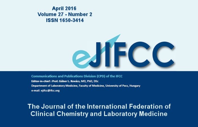
Latest issue of the eJIFCC 2016 Vol 27 no. 2

0

eJIFCC 2016 – Volumen 27 N°2

  • Foreword of the editor. Gábor L. Kovács
  • The Hungarian Society of Laboratory Medicine – serving patients for 70 years. János Kappelmayer
  • Biochemical markers of myocardial damage. Geza S. Bodor
  • Non-invasive assessment of viability in human embryos fertilized in vitro. Gábor L. Kovács, Gergely Montskó, Zita Zrínyi, Nelli Farkas, Ákos Várnagy, József Bódis
  • The clinical value of suPAR levels in autoimmune connective tissue disorders. Barna Vasarhelyi, Gergely Toldi, Attila Balog
  • Deficiencies of the natural anticoagulants – novel clinical laboratory aspects of thrombophilia testing. Zsuzsanna Bereczky, Réka Gindele, Marianna Speker, Judit Kállai
  • Interpretation of blood microbiology results – function of the clinical microbiologist. Katalin Kristóf, Júlia Pongrácz
  • Clinical laboratories – production factories or specialized diagnostic centers. János Kappelmayer, Judit Tóth
  • Adding value in the postanalytical phase. Éva Ajzner
  • Book review — “Patient safety”. Oswald Sonntag

Click here to download.

Agenda


Radio El Microscopio

Ze Xiong

Últimas notas publicadas