Inicio Blog Página 41

The IFCC Foundation for Emerging Nations

0

The IFCC Foundation for Emerging Nations (FEN) is a non-profit making Charitable Trust established in 2016 under Swiss Law by the International Federation of Clinical Chemistry and Laboratory Medicine (IFCC).

The FEN is devoted to fund raising and to supporting programmes that help to improve the quality and delivery of laboratory medicine services, particularly in emerging nations.

A high percentage of all clinical decisions are influenced by the results of laboratory medicine investigations. Consequently, patient safety and clinical outcomes are dependent on the quality of laboratory medicine services.

In emerging nations there is scope for improvement in the quality of laboratory medicine services but educational support is required.

IFCC Members may assist the Foundation for Emerging NationsFEN, in identifying and supporting suitable development projects.

Projects shall be educational in nature and may occur at undergraduate or postgraduate level.

Suitable projects will be in line with the Mission, Aims and Overall Strategic Direction of IFCC, although the FEN is not restricted to supporting projects nominated by IFCC Members.

IFCC has provided ‘start-up’ finance for the FEN. Further sponsorship may be provided by:

  • Other Charitable Trusts active in healthcare
  • Global commercial organisations active in healthcare
  • National or international organisations with an interest in education in health
  • Individual donors interested in improving patient safety in emerging nations

Potential donors to the FEN are encouraged to contact the Chair of the Board of Directors (chair@ifccfoundation.org) to discuss their donation and how it may be used and acknowledged.

Learn more about Foundation for Emerging Nations and visit its website at: www.ifccfoundation.org

Listado de emisiones anteriores

No se encontraron entradas.

Therapeutic anti-CD3 monoclonal antibodies: from bench to bedside

0

Investigations over the last 20 years have shown that anti-CD3 monoclonal antibodies (mAbs) effectively treat autoimmune disease in animal models and have also shown promise in clinical trials. Tolerance induction by anti-CD3 mAbs is related to the induction of Tregs that control pathogenic autoimmune responses. Here, we review preclinical and clinical studies in which intravenous or mucosal administration of anti-CD3 mAbs has been employed and provide an outlook on future developments to enhance the efficacy of this promising therapeutic approach.

Click here to view PDF

Investigating Hyponatremia

0

How is hyponatremia diagnosed?

Labs measure serum sodium with an ion-selective electrode (ISE) using one of two approaches: the indirect method, which involves diluting the sample prior to measurement, and the direct method, which uses neat, undiluted sample. Although newer technologies require much smaller sample volumes, an estimated 83% of laboratories still measure sodium levels using an indirect methodology.

Overall, serum osmolality and urinary sodium measurements are integral to the diagnosis and management of hyponatremia. Also required are clinical assessment and additional laboratory investigations that may include measuring urine osmolality, serum aldosterone, cortisol, and natriuretic peptide levels.

What methods are used to determine serum osmolality?

Serum osmolality is measured using either freezing point depression or vapor pressure techniques. Sodium is the predominant extracellular solute and major contributor to serum osmolality, while other constituents include glucose and urea. Several formulas are available for calculating serum osmolality, but this one is the most widely used: osmolality = 2 x [Sodium] + [Glucose]/18 + [Urea]/2.8.

What does low serum osmolality indicate?

Potential causes of hyponatremia associated with low osmolality (hypotonicity) and excess extracellular fluid include heart failure, liver cirrhosis, and renal impairment. In patients with heart failure, B-type natriuretic peptides will be increased and urinary sodium will be > 20 mmol/L. Urinary sodium will be low in patients with liver cirrhosis, whereas patients with reduced renal function will also exhibit high urinary sodium because the kidneys aren’t properly reabsorbing it.

Hyponatremia, low osmolality, and normal fluid levels can also indicate renal impairment, as well as the syndrome of inappropriate antidiuretic hormone secretion, adrenal insufficiency, and hypothyroidism. Each of these conditions is characterized by normal or low urea, low creatinine and urea/creatinine ratio, low uric acid, and a urinary sodium ≥ 20–30 mmol/L.

Potential causes of hyponatremia, low osmolality, and fluid loss include gastrointestinal disorders, diuretic therapy, and exercise. Another culprit is cerebral salt loss following a subarachnoid hemorrhage, head injury, or neurological procedures. Typical lab values with these conditions include elevated urea, elevated creatinine and urea/creatinine ratio, elevated uric acid, and a reduced urinary sodium < 20–30 mmol/L.

What does normal serum osmolality indicate?

Hyponatremia in patients with normal osmolality (isotonicity) suggests the presence of pseudohyponatremia, for which corrective action is not required. This can be confirmed by testing for elevated lipids and/or proteins.

When lipids and/or proteins are present in plasma in increased concentrations, this can decrease the percentage of plasma water in the blood. This does not impact direct methods for measuring sodium. However, with indirect methods that apply a dilution step prior to sample analysis, this can lead to falsely low sodium results.

What does high serum osmolality indicate?

Hyperglycemia can cause apparent hyponatremia, and may result in osmotic diuresis leading to high osmolality (hypertonicity). High glucose drives water from the intracellular to the extracellular space, diluting sodium concentration. In such a case, measured sodium levels can be corrected by adding 1.6 to 2.4 mmol of sodium for every 100 mg/dL of glucose above 100 mg/dL. High osmolality can also suggest the presence of other solutes such as mannitol.

What leads to a false hyponatremia result?

In addition to pseudohyponatremia, spurious hyponatremia can occur if a sample is contaminated with intravenous fluid containing sodium at less than half that of serum, such as in dextrose infusion. A simple repeat collection often resolves the issue.

Author: Ibrahim A. Hashim, PhD, DABCC, FACB, is the Arthur J. Gill Professor of Pathology, chief of clinical pathology, and clinical chemistry director at the University of Texas Southwestern Medical Center in Dallas.

Source: AACC

Making virus sensors cheap and simple: New method detects single viruses in urine

0

While the technique presently works on just one virus, scientists say it could be adapted to detect a range of viruses that plague humans, including Ebola, Zika and HIV.

“The ultimate goal is to build a cheap, easy-to-use device to take into the field and measure the presence of a virus like Ebola in people on the spot,” says Jeffrey Dick, chemistry graduate student and co-lead author of the study. “While we are still pretty far from this, this work is a leap in the right direction.”

The other co-lead author is Adam Hilterbrand, a microbiology graduate student.

The new method is highly selective, meaning it is only sensitive to one type of virus, filtering out possible false negatives due to other viruses or contaminants.

There are two other commonly used methods for detecting viruses in biological samples, but they have drawbacks: one requires a much higher concentration of viruses and the other requires samples to be purified to remove contaminants. The new method, however, can be used with urine straight from a person or animal.

The other co-authors are Lauren Strawsine, a postdoctoral fellow in chemistry, Jason Upton, an assistant professor of molecular biosciences and Allen Bard, professor of chemistry and director of the Center for Electrochemistry.

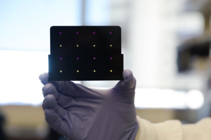
The researchers demonstrated their new technique on a virus that belongs to the same family as the herpes virus, called murine cytomegalovirus (MCMV). To detect individual viruses, the team places an electrode–a wire that conducts electricity, in this case, one that is thinner than a human cell–in a sample of mouse urine. They then add to the urine some special molecules made up of enzymes and antibodies that naturally stick to the virus of interest. When all three stick together and then bump into the electrode, there’s a spike in electric current that can be easily detected.

The researchers say their new method still needs refinement. For example, the electrodes become less sensitive over time because a host of other naturally occurring compounds stick to them, leaving less surface area for viruses to interact with them. To be practical, the process will also need to be engineered into a compact and rugged device that can operate in a range of real world environments.

Source: Sciencedaily.com


Stem cells from diabetic patients coaxed to become insulin-secreting cells

0
Jeffrey R. Millman, PhD, and his colleagues have taken stem cells from the skin of patients with type 1 diabetes and coaxed those cells to differentiate into clusters of insulin-secreting beta islet cells (seen on the computer monitor).

Signaling a potential new approach to treating diabetes, researchers at Washington University School of Medicine in St. Louis and Harvard University have produced insulin-secreting cells from stem cells derived from patients with type 1 diabetes.

People with this form of diabetes can’t make their own insulin and require regular insulin injections to control their blood sugar. The new discovery suggests a personalized treatment approach to diabetes may be on the horizon — one that relies on the patients’ own stem cells to manufacture new cells that make insulin.

The researchers showed that the new cells could produce insulin when they encountered sugar. The scientists tested the cells in culture and in mice, and in both cases found that the cells secreted insulin in response to glucose.

The research is published May 10 in the journal Nature Communications.

“In theory, if we could replace the damaged cells in these individuals with new pancreatic beta cells — whose primary function is to store and release insulin to control blood glucose — patients with type 1 diabetes wouldn’t need insulin shots anymore,” said first author Jeffrey R. Millman, PhD, an assistant professor of medicine and of biomedical engineering at Washington University School of Medicine. “The cells we’ve manufactured sense the presence of glucose and secrete insulin in response. And beta cells do a much better job controlling blood sugar than diabetic patients can.”

Millman, whose laboratory is in the Division of Endocrinology, Metabolism and Lipid Research, began his research while working in the laboratory of Douglas A. Melton, PhD, Howard Hughes Medical Institute investigator and a co-director of Harvard’s Stem Cell Institute. There, Millman had used similar techniques to make beta cells from stem cells derived from people who did not have diabetes. In these new experiments, the beta cells came from tissue taken from the skin of diabetes patients.

“There had been questions about whether we could make these cells from people with type 1 diabetes,” Millman explained. “Some scientists thought that because the tissue would be coming from diabetes patients, there might be defects to prevent us from helping the stem cells differentiate into beta cells. It turns out that’s not the case.”

Millman said more research is needed to make sure that the beta cells made from patient-derived stem cells don’t cause tumors to develop — a problem that has surfaced in some stem cell research — but there has been no evidence of tumors in the mouse studies, even up to a year after the cells were implanted.

He said the stem cell-derived beta cells could be ready for human research in three to five years. At that time, Millman expects the cells would be implanted under the skin of diabetes patients in a minimally invasive surgical procedure that would allow the beta cells access to a patient’s blood supply.

“What we’re envisioning is an outpatient procedure in which some sort of device filled with the cells would be placed just beneath the skin,” he said.

The idea of replacing beta cells isn’t new. More than two decades ago, Washington University researchers Paul E. Lacy, MD, PhD, now deceased, and David W. Scharp, MD, began transplanting such cells into patients with type 1 diabetes. Still today, patients in several clinical trials have been given beta cell transplants with some success. However, those cells come from pancreas tissue provided by organ donors. As with all types of organ donation, the need for islet beta cells for people with type 1 diabetes greatly exceeds their availability.

Millman said that the new technique also could be used in other ways. Since these experiments have proven it’s possible to make beta cells from the tissue of patients with type 1 diabetes, it’s likely the technique also would work in patients with other forms of the disease — including type 2 diabetes, neonatal diabetes and Wolfram syndrome. Then it would be possible to test the effects of diabetes drugs on the beta cells of patients with various forms of the disease.

Source: Washington University School of Medicine in St. Louis

XIVth International Congress of Paediatric Laboratory Medicine (ICPLM)

0

20-22nd October 2017. Durban, South Africa.

On behalf of the Organizing Committee of the XIVth International Congress of Paediatric Laboratory Medicine (ICPLM) and the Task Force on Paediatric Laboratory Medicine (TF-PLM) of the International Federation of Clinical Chemistry and Laboratory Medicine (IFCC), we invite you to the Congress in Durban, South Africa on October 20-22nd 2017. The Congress will be organized in cooperation with the African Federation of Clinical Chemistry. The Congress will focus on the latest scientific and technological achievements in all areas of paediatric clinical and diagnostic laboratory medicine and we are certain that all participants will be enthused by the program. Taking place immediately before the IFCC World Lab Durban 2017, the congress offers you the unique opportunity to gather the latest information in laboratory medicine for children as well as for adult patients.

The scientific program will cover a wide range of topics and includes sessions on genetically determined diseases in children, metabolic disorders, newborn screening, allergy testing, nutrition, endocrinology, paediatric reference intervals, infectious diseases, challenges of the paediatric laboratory, and many other topics. We would also like to encourage you to submit your latest scientific research results to be presented in scientific poster sessions. The program will attract a wide variety of participants including laboratory physicians, pathologists, scientists and technologists, as well as practicing clinicians in paediatrics, neonatology, infectious disease and family medicine.

Durban which is the third largest city in South Africa is a natural paradise known for its gorgeous, safe swimming beaches and subtropical climate, warm Indian ocean, variety of restaurants and rich cultural diversity. Durban is situated on the eastern coast of Africa in Kwazulu Natal province where summer is all year long.

More Information: www.icplm2017.org

No more information is available on infobioquimica.org. For further requests, you can contact the organizers of the event.

IFCC WorldLab Durban 2017

0

Multi-omics and laboratory medicine. 22-25 October 2017, Durban, South Africa. Durban International Convention Center

23rd International Congress of Clinical Chemistry and Laboratory Medicine (IFCC Worldlab 2017)

57th Meeting of the South African Association for Clinical Biochemistry

5th Congress of the African Federation of Clinical Chemistry

Dates to Focus on:

  • 15 May 2017: Poster abstract deadline
  • 15 July 2017: Deadline for reduced fees registration

Main Topics

Autoimmune disease, Allergy, Cardiovascular disease, Bone and joint diseases, Diabetes mellitus and metabolic syndrome, Critical care/emergency medicine, Genetic disease, Endocrinology, Haemostasis, Haematology, Laboratory errors and patient safety, Infectious disease, Liver and gastrointestinal diseases, Laboratory management and information technology, Oncology, Neurological disease, Pharmacogenetics, pharmacogenomics, Paediatric Laboratory Medicine, Pregnancy and neonatology, Point of care testing, Renal function and injury, Quality assessment, standardization, traceability, Toxicology, drugs of abuse, Technology, instrumentation and method evaluation, Vitamins and nutrition, Metabolomics.

Click here for more information 

Updates on the Congress will be availabe at www.durban2017.org

No more information is available on infobioquimica.org. For further requests, you can contact the organizers of the event.

Fasting no longer necessary before cholesterol test, experts say

0

Fasting is a problem for many patients, they explain, and note the latest research shows that cholesterol and triglyceride levels are similar whether people fast or not.

The experts represent the European Atherosclerosis Society (EAS) and the European Federation of Clinical Chemistry and Laboratory Medicine (EFLM) joint consensus initiative.

They refer to new research from Denmark, Canada, and the United States that included over 300,000 people and found it is not necessary to have an empty stomach to check cholesterol levels.

Apart from Denmark, all countries require that patients fast for at least 8 hours before checking their cholesterol and triglyceride levels – referred to as “lipid profile.” In Denmark, non-fasting blood sampling has been in use since 2009.

Arguing their case in a European Heart Journal article, the experts say fasting is a “barrier to population screening” and can be a problem for many patients, particularly children, older adults, diabetes patients, and workers.

“Interestingly, evidence is lacking that fasting is superior to non-fasting when evaluating the lipid profile for cardiovascular risk assessment,” they note. “However, there are advantages to using non-fasting samples rather than fasting samples for measuring the lipid profile.”

Non-fasting and fasting tests ‘not mutually exclusive’

In the journal article, the researchers discuss fasting and non-fasting lipid profile testing and recommends non-fasting for most patients, but they note there are cases when fasting tests are sometimes required, and the two tests should be “viewed as complementary and not mutually exclusive.”

Non-fasting is recommended for most patients, particularly if it is their first lipid profile test, if it is for cardiovascular risk assessment, or if patients express a preference for a non-fasting test.

Non-fasting is also recommended for children, elderly patients, patients on stable drug therapy, and patients admitted with acute coronary syndrome – they will need repeated lipid profile testing later because the condition lowers lipid concentrations.

Diabetic patients should also have non-fasting tests because of higher risk of hypoglycemia, and because fasting can mask high levels of triglycerides (hypertriglyceridemia).

A fasting test can sometimes be required if the non-fasting test shows triglyceride levels above 5 mmol/L (440 mg/dL).

There will also be other specific cases, relating to hypertriglyceridemia, when fasting tests will be necessary. For example, patients with known hypertriglyceridemia being followed in lipid clinic, starting on medications that cause severe hypertriglyceridemia, and recovering from pancreatitis caused by hypertriglyceridemia.

There may also be specific cases where the lab test requires a fasting or morning sample – for example, to test fasting glucose or in monitoring certain drug effects.

However, even in the case of fasting glucose, the authors note that in many countries this measurement is being replaced by measurement of hemoglobin A1c without the need to fast.

Non-fasting testing ‘will improve patient compliance’

The team says that in Denmark, patients, doctors, and testing labs have all benefited from non-fasting testing. It cuts the cost of return visits, emails, phone calls, and follow-up. Doctors also spend less time having to review tests at a later date.

First author Børge Nordestgaard, a professor in the Department of Clinical Medicine at Herlev Hospital, University of Copenhagen, says moving to a system of non-fasting lipid profile testing “will improve patients compliance to preventive treatment aimed at reducing number of heart attacks and strokes, the main killers in the world.”

More patients having the tests also gives doctors opportunity to advise more people on how to prevent heart attacks and strokes, he says. “We hope that non-fasting cholesterol testing will make more patients together with their doctors implement lifestyle changes and if necessary statin treatment to reduce the global burden of cardiovascular disease and premature death.”

Source: Medicalnewstoday.com
 

New microbiome center to combine UChicago, Marine Biological Laboratory, Argonne expertise

0

The University of Chicago, the Marine Biological Laboratory (MBL), and the U.S. Department of Energy’s Argonne National Laboratory announced today a new partnership called The Microbiome Center that will combine the three institutions’ efforts to understand the identity and function of microbes across environments.

These microbial communities–bacteria, viruses and fungi–affect every ecosystem on earth, including human bodies, oceans, our homes, and the land around us.

The new center dovetails with the White House Office of Science and Technology Policy’s National Microbiome Initiative, launched today with the goal of bringing together public and private entities to advance the understanding of microbiome behavior and enable protection and restoration of healthy microbiome function. [More info at http://ow.ly/l7rS300aoKF].

“In the past few years we’ve seen a state change in understanding the roles bacteria play in our world,” said Jack Gilbert who will serve as the Center’s faculty director. Gilbert, a professor in UChicago’s Department of Surgery, has research affiliations at both MBL and Argonne. “This is a unique opportunity to take that knowledge and help drive the next generation of microbiome research forward.”

The Microbiome Center aims to support the research community across the three institutions building on a long tradition of research excellence and collaboration. It will also enable rapid translation to private and clinical sectors, and train a new generation of scientists able to take on fundamental questions about the microbiome, Gilbert said.

“The University of Chicago, MBL and Argonne have already conducted some of the most influential research aimed at understanding and characterizing microorganisms,” said Argonne Director Peter B. Littlewood. “We want to capitalize on this history and expertise in order to advance our capabilities and explore problems that are critical for modern society.”

“This will let us run faster, jump higher, think bigger, and tackle the most important questions facing the field, particular as they impact life in our oceans,” said Marine Biological Laboratory President and Director Huntington Willard.

In the last decade, new techniques–many pioneered at the participating institutions–have allowed researchers to peek at the hidden communities of microbes that populate our world by the trillions.

These studies revealed that microorganisms have complex relationships that affect plants, crops and buildings, and are major players in moving carbon and other elements through massive global cycles. And they live on and in animals and humans, where they both cause and prevent disease–and also regulate some of our most essential functions.

“A greater understanding of microbial communities could affect everything from medicine to agriculture to marine systems and urban development,” Gilbert said.

Microbiome research pulls from many disciplines, including microbiology, immunology, genomics, ecology and evolution, surgery, computation, bioengineering and many more; each institution brings its own expertise to the mix. Argonne has deep expertise in environmental microbiology and sequencing techniques. The Marine Biological Laboratory has extensively studied microbial populations living in oceans, in coastal waterways, and in organisms ranging from marine animals to humans. The University of Chicago is a leader in ecological research, and its medical center has increased its focus on human microbiology and its relation to human health.

Eugene Chang, a professor of biomedical sciences at the University of Chicago who sits on the steering committee for the new Center, highlighted the importance of collaboration to facilitate his research into the role of the microbiome in human health problems, such as obesity and metabolism.

“It really wouldn’t have been possible to answer any of these questions from a single laboratory,” he said. “You could see how each of us look at the same question and each take a different approach. You really needed this multidisciplinary expertise to make discoveries about fundamental principles.”

“It’s really a human need,” said Cathy Pfister, another steering committee member who is a professor in the University of Chicago’s department of ecology and evolution studying ocean microbes. “We’re talking about how ecosystems will manage changes caused by humans–and that will help us understand how we can continue to reap the benefits of the oceans, such as seafood, oxygen and water filtering.”

“The new Center will broaden the look we have at the microbiome,” Willard said. “By asking the same questions on different settings and scales, my guess is that we will discover similar principles at work from people studying inner city microbes and those in deep oceans–and that’s where we can say something fundamental about how life works.”

Source: University of Chicago

Agenda


Radio El Microscopio

Ze Xiong

Últimas notas publicadas