Inicio Blog Página 38

Culture crash: How common pediatric diseases affect the healthy intestinal microbiome

0

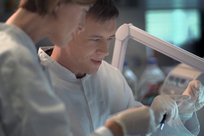
Since the intestinal microbiome is an important regulator of gut health and immune function, Grikscheit and her team investigated how surgical treatment of certain pediatric intestinal diseases have a long-term impact on intestinal flora.

Results of the study, published in the journal Surgery, revealed that for inflammatory and ischemic disorders that require prolonged courses of antibiotics, the microbiome of the intestinal lining remained altered even after the medical condition clinically resolved. Intestine in children who had inflammatory and ischemic disorders had less bacterial diversity and different types of bacteria compared to children who needed intestine surgery that required only a few days of antibiotics such as in the case of obstructive conditions. They also found that patients who underwent intestinal diversion, resulting in digested food passing through just part of their intestine, had little change to their intestinal flora when sites above and below the diversion were compared.

“Our study suggests that the disease process and antibiotic treatment could have a far greater impact on intestinal microbial diversity than surgical intervention,” said Minna Wieck, MD, an investigator and surgical resident at CHLA and first author on the study.

The current study differs from previous reports in that intestinal swabs, which provided a direct method of looking at specific areas of the intestinal mucosa, were studied instead of only relying on stool samples. Also, the researchers used a larger sample size — comparing mucosal bacteria from 43 samples by age, breastfeeding status, disease, acuity and other parameters.

“Alterations in the microbiome are linked to many disease states and may also be associated with increasing surgical complications and long term effects in the children we treat,” said Grikscheit. “Now we need to determine if the disease process or the extended antibiotic regimen is responsible for the changes we identified so that we can effectively care for the medical condition while minimizing any unintended, and potentially, far-reaching effects.”

Source: ScienceDaily

Listado de emisiones anteriores

No se encontraron entradas.

Euromedlab Athens 2017 website is now on-line!

0

22nd IFCC – EFLM European Congress of Clinical Chemistry and Laboratory Medicine
25th Meeting of Balkan Clinical Laboratory Federation
15th National Congress of GSCC-CB

You will find details on the scientific topics as well as the committees, on the sponsorship opportunities as well as the general information.

First Announcement

Dates to focus on:

  • 1 November 2016: Deadline for poster abstract submission
  • 30 April 2017: Deadline for reduced registration fees

Web Page: www.athens2017.org

No more information is available on infobioquimica.org. For further requests, you can contact the organizers of the event.

Vitamin D may not be the great solution to health problems

0

The vital nutrient is widely seen as an important element to good health. Many people place strong belief in its potential benefits in treating a number of medical conditions–such as depression or Multiple Sclerosis–and feel a need to supplement their vitamin D intake. But according to Michael Allan, a professor of Family Medicine and director of Evidence Based Medicine at the University of Alberta’s Faculty of Medicine & Dentistry, much of that belief isn’t validated by science.

“Wouldn’t it be great if there was a single thing that you or I could do to be healthy that was as simple as taking a vitamin, which seems benign, every day? There is an appeal to it. There is a simplicity to it. But for the average person, they don’t need it.” says Allan.

Allan is the lead author of a review published in the Journal of General Internal Medicine that examines the evidence for 10 common beliefs about vitamin D. The beliefs range from the ability of vitamin D to reduce falls and fractures, improve depression and mental well-being, prevent rheumatoid arthritis, treat Multiple Sclerosis, and lessen incidences of cancer and mortality. The review finds little evidence though that supplementation with this vitamin has much of an effect at all.

According to Allan, only a few of the 10 beliefs the team looked into seemed to exhibit some scientific proof. Strongest among them, vitamin D was shown to have a minor impact in reducing the number of falls among the elderly and reducing fractures.

“Even areas that we really thought there was good evidence for benefit early on, don’t seem to be bearing out,” says Allan. “The one that we probably have the most evidence for is fractures. If you were to take a group of people who were at higher risk of breaking a bone–so had about a 15 per cent chance of breaking a bone over the next 10 years–and treated all of them with a reasonable dose of vitamin D for a decade, you’d prevent a fracture in around one in 50 of them over that time.”

“Many people would say taking a drug for 10 years to stop one in every 50 fractures is probably not enough to be meaningful. And that’s the best vitamin D gets as far as we know now.”

Allan says other possible benefits of vitamin D covered in the review were not borne out or are still unproven. He is quick to point out that much of the existing research around vitamin D was poorly executed and consists of poor quality evidence. While he welcomes ongoing research in the area, he says moving forward it needs to consistently be of a higher caliber to be of clinical relevance.

“It makes it really difficult to determine a lot of time if there is anything substantial there that you could tell a patient, ‘You can take this and it can help you this much.’ There’s really not nearly enough there to say that.”

Despite the lack of evidence, belief in the benefits of vitamin D supplementation remains strong. Allan believes much of that stems from misplaced trust in previous research studies showing low vitamin D levels are associated with poor health outcomes, however they don’t prove causation. The bottom line, he says, is that while moderate vitamin D supplementation won’t cause harm to the average healthy person, it won’t heal either.

“The 40 year old person is highly unlikely to benefit from vitamin D,” says Allan. “And when I say highly unlikely, I mean it’s not measurable in present science.”

Source: ScienceDaily

Researchers use liquid biopsy biomarkers to identify prostate cancer before surgery

0

The research, published online today in Nature Communications, advances the quest to develop a precise, non-invasive diagnostic tool that can address the worldwide clinical dilemma of over-treating slow-growing, low-risk prostate cancers in men that may never actually need to be treated, says principal investigator Thomas Kislinger, Senior Scientist at the Princess Margaret Cancer Centre, University Health Network. Dr. Kislinger is also Associate Professor, Department of Medical Biophysics, University of Toronto.

“We believe we have found a better way that allows us to predict which patients have a slow-growing versus aggressive prostate cancer using non-invasive biomarkers. This could eventually help us personalize cancer treatment for these patients.”

Currently, needle biopsies are used to help diagnose prostate cancer. But this technique may not detect hidden tumors, or cancer that has already spread beyond the organ, writes lead author Dr. Yunee Kim: “A fluid-based biomarker would be ideal … to spare patients with indolent (slow-growing) disease from unnecessary procedures, while identifying and treating those who would benefit from treatment intensification.”

The Kislinger team – in collaboration with co-investigators Paul Boutros, Ontario Institute for Cancer Research, and O. John Semmes, Eastern Virginia Medical School, Norfolk – used urine samples containing prostatic secretions from 210 patients after they had undergone digital rectal examinations (DRE). The DRE is the standard clinical “first step” to determine the need for further diagnostic testing of the prostate.

The research took four years and involved samples from 300 patients, says Dr. Kislinger, who specializes in proteomics – the study of the structure and function of proteins involved in the development of cancer.

“We used targeted proteomics to accurately quantify hundreds of proteins in urine samples (post-DRE) to identify liquid biopsy signatures. The first round of research involved 80 patients and quantified 150 proteins that were then narrowed down to 34 for further investigation. The next round involved a second, independent cohort of 210 patients.

“Applying computational biology, we used the quantitative data from mass spectrometry to develop the fluid biomarkers for aggressive prostate cancer.”

Dr. Kislinger says: “The next step will be further studies with urine samples from 1,000 international patients to validate if the biomarkers identified have broader clinical utilities in prostate cancer.”

Source: sciencenewsline.com

IFCC’s eNews May – June 2016 is now available!

0

The issue opens with an article in memoriam of Prof. Siest, IFCC Past President. News of IFCC initiatives and accomplishments in this edition include a report on the Biomarkers on Alzheimer’s disease meeting, the launch of the IFCC eNews Flash, the IFCC Medal, and a focus on Shaping the Future of Laboratory Medicine. Other news items from IFCC Federations and National Associations allow the reader to keep up with current interesting activities around the laboratory medicine world.

Test holds potential to diagnose myriad conditions with drop of blood

0

The results, reported in the Journal of Immunological Methods and funded by the Bill & Melinda Gates Foundation, are the first evidence that it is possible to develop blood tests for any infectious disease by screening random libraries of non-biological molecular shapes.

“This ‘needle-in-a-molecular haystack’ approach is a new way to develop diagnostic assays,” said senior author Donald S. Burke, M.D., Pitt Graduate School of Public Health dean and director of Pitt’s Center for Vaccine Research. “The method does not rely on starting with known viral components. This is important because there are conditions for which there isn’t a known antigen, such as newly emerged epidemics, autoimmune diseases or even responses to traumatic injury.”

When a person’s immune system is faced with an antigen or foreign invader, such as an infectious disease, or even an injury with tissue damage, it responds by producing antibodies. Like puzzle pieces, specific parts of the surface of these antibodies fit to the shape of the molecules on the invader or the damaged tissue.

The Pitt researchers used a technique pioneered by co-author Thomas Kodadek, Ph.D., of the Scripps Research Institute, that synthesizes random molecular shapes called “peptoids” hooked onto microscopic plastic beads. The technique can produce millions of molecular shapes. The peptoids are not organic, but if they match to the corresponding shape on an antibody, that antibody will connect to them, allowing the scientist to pull out that bead and examine that peptoid and its corresponding antibody.

Using this technique, Dr. Burke’s team chemically generated a huge library of random molecular shapes. Then, using blood from HIV-infected patients and from non-infected people, the researchers screened a million of these random molecular shapes to find the ones that bound only to antibodies present in the blood of HIV-infected patients, but not the healthy controls. No HIV proteins or structures were used to construct or select the peptoids, but the approach, nonetheless, successfully led to selection of the best molecular shapes to use in screening for HIV antibodies.

The team then resynthesized that HIV-antibody-targeting peptoid in mass and tested it by screening hundreds of samples from the Multicenter AIDS Cohort Study (MACS), a confidential research study of the natural history of treated and untreated HIV/AIDS in men who have sex with men (supported by the National Institutes of Health). Study co-author Charles Rinaldo, Ph.D., chair of Pitt Public Health’s Department of Infectious Diseases and Microbiology and director of the Pittsburgh arm of the MACS, selected the samples, but blinded the testers to which samples were HIV-positive or -negative. The test distinguished between the samples of HIV-positive blood and HIV-negative blood with a high degree of accuracy.

“This technology means that we may be able to take a single drop of blood from a patient and detect antibodies to all manner of infections, cancers or other conditions they may be carrying or been exposed to. We hope that this is the first step toward development of an ‘Epi-chip’ that can be used to reconstruct a person’s entire exposure history,” said Dr. Burke, who also holds the UPMC-Jonas Salk Chair of Global Health at Pitt.

Source: Eurekalert


Study reveals impact of antibiotic treatment, other factors on the infant gut microbiome

0

A comprehensive analysis of changes in the intestinal microbial population during the first three years of life has revealed some of the impacts of factors such as mode of birth – vaginal versus cesarean section – and antibiotic exposure, including the effects of multiple antibiotic treatments. In the June 15 issue of Science Translational Medicine, the team led by investigators from Massachusetts General Hospital (MGH) and the Broad Institute describes findings that may help understand how the gut microbiome is established and how the combination of microbes in individual children may contribute to the risk of developing conditions like type 1 diabetes and inflammatory bowel disease.

“One of the key motivations of microbiome research is that the microbial population of early childhood appears to be critical to human health, in that decreased diversity of the gut microbiome has been implicated in a number of allergic and autoimmune diseases,” says Ramnik Xavier, MD, PhD, chief of the MGH Gastrointestinal Unit and an institute member at the Broad. “Not only did our study analyze the gut microbiome at a high resolution that allowed us to identify both microbial species and strains, but by following our study participants over time we also were able to uncover findings that would not have been revealed by single samples from each patient.

In collaboration with a team of Finnish researchers they have worked with for several years, the MGH/Broad team enrolled a group of 39 children from whom monthly stool samples were taken beginning after birth and continuing until age 36 months. Each sample was analyzed with a standard, RNA-based sequencing procedure used to identify microbial populations, and more detailed whole-genome sequencing was conducted on about 25 percent of samples to reveal the specific strains of identified microbial species. During the study period, 20 of the participants received antibiotics to treat respiratory or ear infections, with those children receiving from 9 to 15 treatments.

Many features of the developing gut microbiome were found to be consistent across all study participants, with the presence and abundance of particular species rising and falling at similar age points. The researchers also found several clear differences from the findings of previous studies regarding the impact of breastfeeding. Earlier studies comparing breast-fed with formula-fed children have reported increased abundance of Bifidobacterium species in those who were breast-fed for longer periods of time. All of the children in this study were breastfed for some period of time, and while there was some correlation between the length of breastfeeding and levels of Bifidobacteria, some of the children in this group had low levels of those bacteria even while being breastfed.

Previous studies also have reported finding a particular microbiome signature, with low abundance of the Bacteriodes genus, in cesarean-section-delivered children during the first 6 months of life. In the current study, the researchers found the same pattern in the four cesarean children but were surprised to find it also occurred in seven of the vaginally born children. No identified factors, including maternal antibiotic treatment, differentiated between vaginally born children with or without the low-Bacteriodes signature, but since this pattern has been associated with reduced overall diversity of the microbiome, it bears further investigation, Xavier notes.

Children who had been exposed to antibiotic treatment had a reduction in the diversity of their microbial population, a difference that was even greater in those who also had the low-Bacteriodes signature. Whole-gene sequencing also found that, in antibiotic-exposed children, bacterial species tended to be fewer and dominated by a single strain, instead of the several species and strains seen in those not treated with antibiotics. The analysis of many samples taken over time revealed that the microbiomes of antibiotic-exposed children were less stable, particularly around the time of antibiotic treatment.

The presence of genes known to confer antibiotic resistance rose rapidly during antibiotic treatment. Levels of resistance genes encoded on microbial chromosomes dropped quickly after treatment was discontinued. But resistance genes encoded on small DNA molecules called mobile elements – one means by which resistance genes can be transmitted among bacteria – persisted much longer after antibiotic withdrawal.

“Some of the things we’d like to investigate next are how the microbiome gets established during the first week of life – particularly what the primary mechanisms of transmission are – how the composition of the early-life gut microbiome affects children’s health, and what factors underlie the resilience of the infant microbiome, says Xavier, who is the Isselbacher Professor of Medicine in Gastroenterology at Harvard Medical School and a member of the MGH Center for Computational and Integrative Biology (CCIB). “The kind of high-resolution sequencing done in this study should lead to better understanding of the natural history of the infant gut microbiome and the impact of perturbations caused by antibiotics and environmental factors.”

Source: massgeneral.org


A new biomarker for nerve cell damage

0

Scientists at the German Center for Neurodegenerative Diseases, the Hertie Institute for Clinical Brain Research and the University of Tübingen have identified proteins in the blood and cerebrospinal fluid that reflect nerve cell damage.

The results of the study, published in the journal Neuron, suggest that the concentration of these ‘neurofilament light chain proteins’ could provide information about the progression of neurodegenerative diseases and the effects of treatment. Such a biomarker would be valuable for developing therapies.

“The results of our research indicate that disease progression can be tracked by monitoring the concentration of neurofilament light chain proteins. According to our study this is possible in both animal models and humans,” says Mathias Jucker, who leads a research group at the DZNE’s Tübingen site and is also a director of the Hertie Institute. “We have also found that the measured levels react sensitively when pathological hallmarks in the brain are influenced experimentally. It may thus be possible to evaluate the effect of a treatment by measuring the concentration of light neurofilaments, both in preclinical laboratory studies and in clinical trials. Such a biomarker would be a major benefit for developing treatments.”

Parts of the cytoskeleton

“Neurofilament light chain proteins” are parts of the cytoskeleton that gives nerve cells shape and stability. These thread-like molecules are therefore mainly located inside cells, but may be released due to damage.

Professor Jucker and his colleagues – including Mehtap Bacioglu, first author of the current publication – took this known fact as the basis for investigating the concentration of neurofilament light chains proteins in the blood and cerebrospinal fluid (liquor). For this, they looked at mice that showed the typical features of neurodegenerative diseases, namely deposits of aggregated “alpha-synuclein”, “tau” or “beta-amyloid” protein in their brains. Such deposits are associated with nerve cell damage. Besides, the scientists examined biomaterials from patients with Alzheimer’s, Parkinson’s and other neurodegenerative diseases.

Sensitive measurements

In mice, a close association was found between the concentration of neurofilament proteins in the liquor and blood. Moreover, the more advanced the brain damage, the higher the measured levels. If the neurological lesions were induced or inhibited protein levels increased or dropped accordingly. In patients, blood and liquor readings also correlated strongly. Furthermore, levels were higher than in healthy people.

A tool for developing treatments

“The special potential of this biomarker comes from the fact that it is significant in both animals and humans. Therefore, the results from animal models can be translated into clinical studies and their findings may be directly compared. This is critical for the development of new treatments,” says Jucker. “What’s more, we don’t have to rely on withdrawals of liquor. The lumbar puncture required to obtain cerebrospinal fluid can be stressful for the person undergoing it. Our study shows that blood levels also provide information about neurodegeneration in the brain, because the concentrations of the neurofilaments in the blood and cerebrospinal fluid are closely coupled. A simple blood sample may therefore be sufficient when performing clinical studies on humans.”

Source: uni-tuebingen.de


Scientists discover biomarkers that could give cancer patients better survival estimates

0

People with cancer are often told by their doctors approximately how long they have to live, and how well they will respond to treatments, but what if there were a way to improve the accuracy of doctors’ predictions?

A new method developed by UCLA scientists could eventually lead to a way to do just that, using data about patients’ genetic sequences to produce more reliable projections for survival time and how they might respond to possible treatments. The technique is an innovative way of using biomedical big data — which gleans patterns and trends from massive amounts of patient information — to achieve precision medicine — giving doctors the ability to better tailor their care for each individual patient.

The approach is likely to enable doctors to give more accurate predictions for people with many types of cancers. In this research, the UCLA scientists studied cancers of the breast, brain (glioblastoma multiforme, a highly malignant and aggressive form; and lower grade glioma, a less aggressive version), lung, ovary and kidney.

In addition, it may allow scientists to analyze people’s genetic sequences and determine which are lethal and which are harmless.

The new method analyzes various gene isoforms — combinations of genetic sequences that can produce an enormous variety of RNAs and proteins from a single gene — using data from RNA molecules in cancer specimens. That process, called RNA sequencing, or RNA-seq, reveals the presence and quantity of RNA molecules in a biological sample. In the method developed at UCLA, scientists analyzed the ratios of slightly different genetic sequences within the isoforms, enabling them to detect important but subtle differences in the genetic sequences. In contrast, the conventional analysis aggregates all of the isoforms together, meaning that the technique misses important differences within the isoforms.

SURVIV (for “survival analysis of mRNA isoform variation”) is the first statistical method for conducting survival analysis on isoforms using RNA-seq data, said senior author Yi Xing, a UCLA associate professor of microbiology, immunology and molecular genetics. The research is published today in the journal Nature Communications.

The researchers report having identified some 200 isoforms that are associated with survival time for people with breast cancer; some predict longer survival times, others are linked to shorter times. Armed with that knowledge, scientists might eventually be able to target the isoforms associated with shorter survival times in order to suppress them and fight disease, Xing said.

The researchers evaluated the performance of survival predictors using a metric called C-index and found that across the six different types of cancer they analyzed, their isoform-based predictions performed consistently better than the conventional gene-based predictions.

The result was surprising because it suggests, contrary to conventional wisdom, that isoform ratios provide a more robust molecular signature of cancer patients than overall gene abundance, said Xing, director of UCLA’s bioinformatics doctoral program and a member of the UCLA Institute for Quantitative and Computational Biosciences.

“Our finding suggests that isoform ratios provide a more robust molecular signature of cancer patients in large-scale RNA-seq datasets,” he said.

The researchers studied tissues from 2,684 people with cancer whose samples were part of the National Institutes of Health’s Cancer Genome Atlas, and they spent more than two years developing the algorithm for SURVIV.

According to Xing, a human gene typically produces seven to 10 isoforms.

“In cancer, sometimes a single gene produces two isoforms, one of which promotes metastasis and one of which represses metastasis,” he said, adding that understanding the differences between the two is extremely important in combatting cancer.

“We have just scratched the surface,” Xing said. “We will apply the method to much larger data sets, and we expect to learn a lot more.”

Source: Newsroom UCLA

Agenda


Radio El Microscopio

Ze Xiong

Últimas notas publicadas