Inicio Blog Página 31

Discovery of Rapidly Adapting Antibody Opens Door for Universal Flu Vaccines

0
In a newly published study, scientists report that they have discovered a type of immune antibody that can rapidly evolve to neutralize a wide array of influenza virus strains—including those the body hasn’t yet encountered. [extender01/Getty Images]

Now, it would seem, investigators at the Dana-Farber Cancer Institute are looking to take the lead in this critical discussion because they have recently published data on the discovery of a new type of immune antibody that can evolve rapidly to neutralize a broad range of influenza virus strains—including those the body hasn’t yet encountered. The revelation of the body’s ability to make the adaptable antibody suggests potential strategies for creating improved or even universal influenza vaccines.

The new antibody, named mAb 3I14, is a broadly neutralizing antibody (bnAb), so-called because it can recognize and disable a diverse group of the 18 different strains of influenza virus that circulate the globe. The Dana-Farber team reported that the 3I14 antibody demonstrated it could efficiently neutralize the two main types of influenza A virus—groups 1 and 2—and protected mice against lethal doses of the virus.

“We report a haemagglutinin (HA) stem-directed bnAb, 3I14, isolated from human memory B cells, that utilizes a heavy chain encoded by the IGHV3-30 germline gene,” the authors wrote. “MAb 3I14 binds and neutralizes groups 1 and 2 influenza A viruses and protects mice from lethal challenge. Analysis of variable regions of VH [heavy-chain] and VL [light-chain] germline back-mutants reveals binding to H3 and H1 but not H5, which supports the critical role of somatic hypermutation in broadening the bnAb response.”

The findings from this study were published recently in Nature Communications in an article entitled “A Broadly Neutralizing Anti-Influenza Antibody Reveals Ongoing Capacity of Haemagglutinin-Specific Memory B Cells to Evolve.”

The 3I14 antibody is made by the human immune system’s memory B cells—immune cells that circulate in the blood and reside within the spleen and bone marrow. When an individual is exposed to an infectious agent or receives a vaccine made from pieces of that agent, B cells respond to the invaders and can generate a memory of the particular type or strain. Pools of these memory B cells constitute a reserve defensive force that can quickly recognize and attack the microbe or virus should it enter the body again.

Unfortunately, influenza’s hypermutability makes it difficult to protect against one-time vaccination strategies. Viral changes occur every flu season and are responsible for the seasonal flu that we are vaccinated against yearly. The more dramatic changes that occur when new viruses emerge from animal and bird reservoirs are responsible for potentially more severe pandemics, such as the deadly H1N1 strain that emerged in 2009.

The discovery of the new bnAbs came after senior study investigator Wayne Marasco, M.D., Ph.D., a cancer immunologist and virologist at Dana-Farber, along with his colleagues took blood samples from seven blood bank donors that were shown to harbor these types of antibodies and challenged their immune B cells in the laboratory with an array of flu viruses. The researchers ultimately identified one B-cell population “that recognized all the strains we screened against it,” Dr. Marasco said. Sorting through the B cells’ DNA, the researchers isolated the gene that carried the instructions for creating the 3I14 antibody.

The research team found that the antibody could bind to the unchanging stem portion of flu viruses. Moreover, they reported that the antibody’s genetic makeup gave it the flexibility to adapt or evolve, through mutations, to neutralize a myriad of flu viruses.

To test the antibodies’ full capabilities, the investigators challenged the B cells with a bird flu virus of the H5 type that the immune cells had never encountered. Although the 3I14 antibody didn’t initially bind strongly to the virus, the researchers introduced a single DNA mutation that increased its binding strength to H5 by ten times. “To our delight, we made one mutation, and it did the job,” Dr. Marasco noted. “This was a simple mutation that would readily occur in nature.”

“These data provide evidence that memory B cell evolution can expand the HA subtype specificity,” the authors concluded. “Our results further suggest that establishing an optimized memory B cell pool should be an aim of ‘universal’ influenza vaccine strategies.”

Source: GEN

Listado de emisiones anteriores

No se encontraron entradas.

Gonorrhea May Become Resistant to All Antibiotics Sooner Than Anticipated

0

Experts have been worried for a while that time is running out for the last working cures for gonorrhea infections. Some revealed there may be even less time left on the clock than had been previously thought.

Scientists from the Centers for Disease Control and Prevention and Hawaii’s department of health reported a cluster of cases of gonorrhea in that state in which the Neisseria gonorrhoeae bacteria showed high-level resistance to one of the drugs, azithromycin, as well as reduced susceptibility to the second drug, ceftriaxone.

The two drugs are used in combination, a move experts hope will slow down the bacteria’s relentless march through the antibiotic medicine cabinet.

There had already been signs that the bacteria, which have vanquished multiple other antibiotics, were starting to be able to evade these ones as well. Since 2005, there have been four isolated cases reported in which the Neisseria gonorrhoeae bacteria had reduced susceptibility to both drugs. But this is the first time a cluster of such cases has been seen in the US.

“Our last line of defense against gonorrhea is weakening,” Dr. Jonathan Mermin, director of CDC’s National Center for HIV/AIDS, Viral Hepatitis, STD, and TB Prevention said in a statement. “If resistance continues to increase and spread, current treatment will ultimately fail and 800,000 Americans a year will be at risk for untreatable gonorrhea.”

Untreated gonorrhea can cause chronic pelvic pain, infertility, and ectopic pregnancies—in which a fertilized egg starts to develop in a fallopian tube, not the uterus. Ectopic pregnancies can be life-threatening.

The cluster of cases described by Dr. Alan Katz of Hawaii’s state board of health involved six men and a woman, but was in fact undoubtedly larger. They did not have sex with each other, so must have been infected by other people. Katz said the seven people reported a total of eight sexual partners. Four of the eight agreed to be tested, but none was infected.

Analysis of the genetic sequences showed they were closely related, Katz said.

All seven people were cured of their infections. But it seems only a matter of time before treatment failures occur. Earlier this year doctors from Britain reported that a man infected with gonorrhea was not cured by his first course of antibiotics, though follow-up treatment three months later did quell the infection.

If this combination of drugs fail, there is no other treatment option currently available. Several experimental antibiotics are in development and one has recently been shown to be effective in a Phase 2 trial. But it is still likely years away from hitting the market.

Source: Scientific American

Rapid Influenza Testing: Three Key Questions for Laboratorians

0

These pertinent and timely questions will remain so as influenza viruses keep their status as a top driver of respiratory illness with potentially severe consequences in immuno-suppressed or compromised individuals.

A number of different techniques are available to diagnose influenza, including viral cell culture, direct fluorescent antigen (DFA), rapid influenza diagnostic tests (RIDT), and molecular methods. Since appropriate cohorting and treating of patients with influenza depends on accurate and timely diagnosis, many hospitals and laboratories have opted for point-of-care or in-laboratory rapid diagnostic testing methods, including RIDTs or molecular methods.

Many Food and Drug Administration (FDA)-approved RIDTs detect influenza A and/or B. These methods are quick (<30 minutes), easy to perform in either physicians’ offices or laboratories, and specific. However, they lack sensitivity (50%-70%) compared to viral culture. The primary FDA-approved rapid molecular testing methods use either reverse-transcription polymerase chain reaction (RT-PCR) or loop-mediated isothermal amplification (LAMP) technologies.

Molecular methods are much more sensitive than RIDTs, but depending on the assay, compromise slightly on turnaround time, cost, and ease of use. Molecular methods also usually detect both influenza A and B together, and often subtype the influenza A viruses. Clinical laboratories handle the majority of molecular testing as opposed to doctors’ offices or inpatient units. However, this is now changing with the availability of LAMP testing, which has been designed to be faster than RT-PCR and used in a point-of-care testing environment.

Regardless of method used and site of sample collection or testing, in my experience, the following are three of the most common influenza testing questions clinical laboratories handle during flu season.

What specimen type should I collect, and how should it be collected?

For any rapid influenza assay, appropriate specimen collection and handling is key to accurate diagnosis. While FDA-approved specimen types vary by assay, nasopharyngeal swabs and nasal washes/aspirates are the most sensitive for detecting influenza virus. Throat and nasal swabs are FDA-approved for certain, but not all, rapid diagnostic tests, and they typically have inferior recovery of the virus.

Requests for testing other, non-FDA approved sample types such as bronchoalveolar lavage samples or endotracheal aspirates are handled differently depending on the testing laboratory. These specimen types require a separate validation or a disclaimer if being tested. Some laboratories may prefer to send these requests to a reference laboratory that has validated these specimen types.

The specific swabs to be used for specimen collection vary depending on the rapid assay performed. Some assays may provide or recommend a specific swab for specimen collection. I can’t overstate the importance of using the recommended swabs for each assay, as certain swab materials can be inhibitory to certain assays.

To preserve their integrity, specimens should be placed in the appropriate, assay-specific, universal or viral transport medium and sent to the testing laboratory immediately or refrigerated if a transport delay is expected. Inappropriate sample types include swabs placed in dry containers or any transport medium other than those listed in the manufacturer’s instructions for the assay.

How quickly will I get my patient’s test result?

The answer to this question depends on the type of test being performed, where it is being performed, and which turnaround times hospitals and laboratories have agreed upon. RIDTs and LAMP assays have the fastest turnaround times (<30 minutes), whereas RT-PCR assays generally take 1–1.5 hours. In reality, turnaround times can be much longer depending on how quickly the specimen makes it to the lab, laboratory staffing, and whether or not the assay is a part of a lab’s STAT testing menu.

Is the reported result reliable?

Since the performance characteristics of RIDTs and molecular methods differ dramatically, appropriate patient management depends on proper interpretation of test results. Due to the low-to-moderate sensitivity of RIDTs, false negatives are common, especially during peak influenza season. Negative results do not exclude influenza infection, and providers should consider another diagnostic method—DFA, culture, or molecular—for confirmation if clinical suspicion of influenza is high. Due to the high specificity of most RIDTs, a positive result is considered reliable. Molecular methods, on the other hand, are both sensitive and specific, with both negative and positive results typically reliable. However, an important limitation of molecular methods is the possibility of detecting non-viable or non-replicating virus.

In addition to the performance characteristics of the specific test used, several other factors influence reliability of rapid test results, including specimen type, specimen collection and handling, and how soon after symptom onset the specimen was collected—ideally between 12 hours and 3 days afterwards. Assay-specific manufacturers’ recommendations should be followed to minimize the possibility of inaccurate results.

Armed with answers to these questions, labs will be ready to go when flu season strikes.

Author: Tanis Dingle, PhD, D(ABMM), FCCM.

Source: AACC

Should All Children and Teens Be Screened For High Cholesterol?

0

The guideline applies to youths with no known risk factors for heart disease. Screening with a lipid profile is recommended for those who have risk factors, such as diabetes, high blood pressure, or being overweight.

In contrast, the 2011 guidelines from the National Cholesterol Education Program (NCEP), then a division of the National Institutes of Health, recommended routine cholesterol screening for all children and young adults. Specifically, they recommended that all children between the ages of 9 and 11 be screened once and then have additional lipid testing between the ages of 17 and 21. The American Academy of Pediatrics (AAP) endorses recommendations for universal screening.

The USPSTF evaluated this topic because of a concern that youths with high cholesterol are more likely to have high cholesterol levels as adults, which can lead to an increased risk of heart attacks and strokes.

To reach its decision, the Task Force looked at research studies conducted on high cholesterol in children caused by genetics, or family history, as well as high cholesterol caused by a combination of factors, such as a high fat diet. The data available from current studies are not sufficient to recommend for or against routine screening, concluded the Task Force, because:

  • The evidence showed it was difficult to predict which children with high cholesterol would continue to have the condition as adults.
  • The evidence did not indicate whether testing for and treating high cholesterol in children and young adults would lead to better cardiac health when those children became adults.
  • There is a lack of evidence on potential harms from the medications used to treat high cholesterol in children and teens.

“By issuing an “I” (for “insufficient”) statement, we are calling for more research to better understand the benefits and harms of screening and treatment of lipid (high cholesterol and triglycerides) disorders in children and teens and on the impact these interventions may have on their cardiovascular health as adults,” said David Grossman, M.D., M.P.H, the vice chair of the Task Force. Dr. Grossman is also a professor of health services and an adjunct professor of pediatrics at the University of Washington.

The AAP, which currently endorses universal screening, is evaluating the current research and could release an updated statement in a year or two, says Steven Daniels, MD, PHD, chair of the department of pediatrics at the University of Colorado School of Medicine as well as chair of the AAP’s committee on nutrition.

Meanwhile, parents of children and teens who have questions should have a conversation with their healthcare practitioners to help make decisions about cholesterol testing.

“In the absence of evidence, health care professionals should continue to take each patient’s individual risks and circumstances in consideration, and use their best judgment when deciding whether or not to screen,” said Dr. Grossman.

Source: Lab Tests Online

Mysterious New Mosquito-Borne Disease Found in Caribbean

0
Researchers have identified a patient in Haiti with a serious mosquito-borne illness that has never before been reported in the Caribbean nation. [mycteria/Getty Images]

Known as Mayaro virus, this pathogen belongs to the Alphavirus genus, is closely related to chikungunya, and produces a dengue-like illness that is often accompanied by long-lasting arthralgia (joint pain). Whether this case signals the start of a new outbreak in the Caribbean region or is an isolated incident is currently unknown.

The findings from the new report were published online in Emerging Infectious Diseases in an article entitled “Mayaro Virus in Child with Acute Febrile Illness, Haiti, 2015.”

“While current attention has been focused on the Zika virus, the finding of yet another mosquito-borne virus that may be starting to circulate in the Caribbean is of concern,” explained senior study investigator Glenn Morris, M.D., director of the UF Emerging Pathogens Institute (EPI). “Hopefully we will not see the same massive epidemics that we saw with chikungunya, dengue, and now Zika. However, these findings underscore the fact that there are additional viruses ‘waiting in the wings’ that may pose threats in the future, and for which we need to be watching.”

The Mayaro virus case was identified from a blood sample taken in January 2015 from an 8-year-old boy in rural Haiti. The patient had a fever and abdominal pain, but no rash or conjunctivitis. Since faculty from the UF Emerging Pathogens Institute were in the region during and after the 2014 chikungunya outbreak, plasma samples were obtained from febrile children and analyzed for the presence of chikungunya virus RNA using the genetic identification technique reverse transcription polymerase chain reaction, RT-PCR.

“Mayaro virus (MAYV) is a single-stranded positive RNA virus that was first isolated in Trinidad in 1954 and is one of the viruses that comprise the Semliki Forest virus complex,” the authors wrote. “Its transmission cycle is thought to occur mainly through mosquito vectors, especially those of genus Haemagogus, but Aedes spp. mosquitoes may also be competent vectors. The natural reservoirs of MAYV have been reported to be sylvatic (wild) vertebrates, mainly nonhuman primates but also birds and reptiles.”

The plasma samples were examined by UF researchers, in Gressier, Haiti, and were then sent to EPI for additional virology and molecular analyses, focusing on the detection of chikungunya, dengue, and Zika viruses. Interestingly, dengue virus was detected in the patient, in addition to a “new” virus —which was subsequently identified as Mayaro.

“The virus we detected is genetically different from the ones that have been described recently in Brazil, and we don’t know yet if it is unique to Haiti or if it is a recombinant strain from different types of Mayaro viruses,” concluded lead study author John Lednicky, Ph.D., an associate professor in the environmental and global health department at the UF College of Public Health and Health Professions.

The UF team and infectious disease agencies are working closely to keeping an eye on the situation in Haiti and other Caribbean nations to identify and potentially get out in front of any emerging outbreaks.

Source: GEN

Circulating Immune Cells as Biomarkers for Idiopathic Pulmonary Fibrosis

0
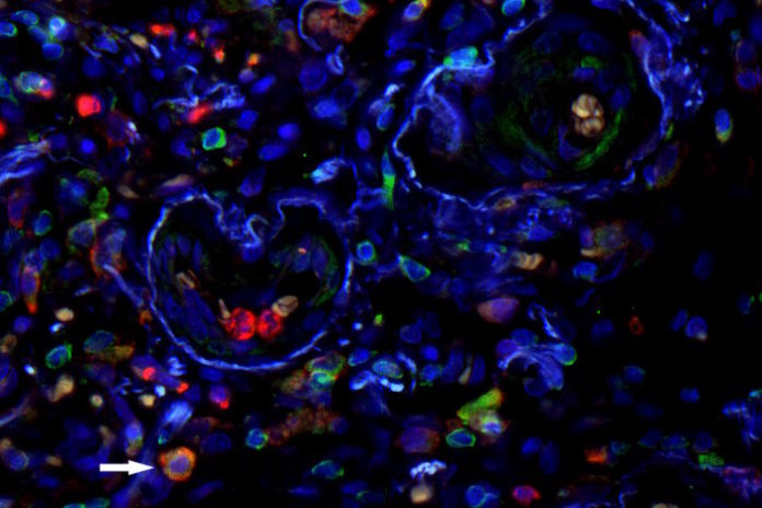
Staining of cells in lung tissue: the surface molecules CD11 and CD33 are shown in green and red, respectively. Nuclei are shown in blue. MDSC are positive for both surface markers and consequently appear orange (arrow). Source: Helmholtz Zentrum München

Patients with fibrotic lung diseases*, such as idiopathic pulmonary fibrosis (IPF), show progressive worsening of lung function with increased shortness of breath and dry cough. To-date, this process is irreversible, which is why scientists are searching for novel biomarkers or indicators, which enable earlier diagnosis of this disease, with the aim to better interfere with disease progression.

A team of scientists at the Comprehensive Pneumology Center (CPC) at Helmholtz Zentrum München headed by Professor Oliver Eickelberg, Chairman of the CPC and Director of the Institute of Lung Biology as well as the DZL at the Munich partner site, have now discovered that myeloid-derived suppressor cells (MDSC) may serve as such biomarkers. “The role of MDSC has been most extensively studied in cancer, where they suppress the immune system and contribute to a poor prognosis,” explained first author Isis Fernandez, MD. The current study suggests that similar mechanisms are also at work in IPF.

In collaboration with the Department of Internal Medicine V (Director: Professor Jürgen Behr) of the Munich University Hospital, the team examined blood samples of 170 study participants, including 69 IPF patients, in terms of the composition of circulating immune cell types. In each patient, these were correlated with lung function. Strikingly, the MDSC count in IPF patients was significantly higher than in the healthy control group. At the same time, the researchers observed that there was an inverse correlation between lung function and circulating MDSC counts: the poorer the lung function, the higher the MDSC count. In control groups of patients with chronic obstructive pulmonary disease (COPD) or other interstitial lung diseases, this inverse correlation was not found. “We conclude that the number of MDSC reflects the course of the disease, especially in IPF,” said Fernandez.

To obtain an indication of whether the cells themselves could be the cause of the deterioration in lung function, the researchers measured the activity of genes that are typically expressed by immune cells. They found that these genes were expressed less frequently in samples that exhibited high MDSC counts. This indicates that MDSC – similar to their role in cancer – also compromise the immune system in IPF, according to the scientists.

A look into the lung tissue of IPF patients supports this assumption. “We were able to show that MDSC are primarily found in fibrotic niches of IPF lungs characterized by increased interstitial tissue and scarring, that is, in regions where the disease is very pronounced,” said Eickelberg. “As a next step, we seek to investigate whether the presence of MDSC can serve as a biomarker to detect IPF and to determine how pronounced it is.” In addition, the researchers want to investigate the mechanisms of accumulation in more detail. “Controlling accumulation or expansion of MDSC or blocking their suppressive functions may represent a promising treatment options for patients with IPF,” said Eickelberg.

Sourcehelmholtz-muenchen.de

A role for siderophores in Klebsiella pneumoniae pathogenesis

0

The bacterium, estimated to be the third most common cause of hospital-acquired infections in the United States in a recent study, can cause a wide range of infections such as pneumonia, bacteremia, wound or surgical site infections and urinary tract infections. What’s more, Klebsiella pneumoniae is rapidly becoming resistant to all known antibiotics; resistant forms are considered an urgent threat to public health by the Centers for Disease Control and Prevention.

What scientists have known about the organism is that it secretes small molecules called siderophores that enable it to acquire iron from a host and fuel its spread. Siderophores are thought to worsen infection by promoting bacterial growth. But a new study in mBio this week demonstrates that siderophores do even more to help Klebsiella invade.

In mice infected with K. pneumoniae, siderophores caused the animals to turn on proteins that increase inflammation and activated a master protein called HIF-1 alpha that paved the way for bacteria to spread from the lungs to the spleen. HIF-1 alpha controls the ability of substances to pass through blood vessels and inflammatory gene expression.

“We’ve known for a long time that siderophores are critical for bacteria to cause infection, because they steal iron from the host to grow,” says senior study author Michael Bachman, M.D., Ph.D., assistant professor of clinical pathology and associate director of the Clinical Microbiology Laboratory at the University of Michigan in Ann Arbor. “This study sheds some light on the consequences of that. When the bacterium steals this iron, what’s likely happening is the host cells are becoming stressed and are inducing inflammation and cell signaling pathways that actually worsen the infection by allowing the bacteria to escape from the lungs to the spleen.”

A barrier to studying siderophores in vivo in the past, Bachman says, is that they allow bacterial proliferation, which in turn may increase inflammation, tissue damage and bacterial dissemination. “What we tried to do was control for levels of bacteria, and then compare infections in which the Klebsiella made siderophores and in which it didn’t,” he explains. To study the effects of siderophore secretion independent of increased bacterial growth, Bachman’s team employed tonB mutants capable of disrupting normal use of siderophores, so the organism could secrete siderophores but not use them.

The investigators infected mice with either a strain of K. pneumoniae that makes three siderophores called enterobactin (Ent), salmochelin (Sal) and yersiniabactin (Ybt), or a mutant form of the bacterium that could not produce siderophores. Mice infected with the siderophore-producing strain had more bacteria invading from the lung to the spleen and they activated the cytokines IL-6, CXCL1 and CXCL2.

“We had assumed that siderophores simply helped the bacteria grow more, triggering more inflammation and spread,” Bachman says. “Now we know siderophores can trigger these effects directly.”

Additional laboratory experiments in the mice revealed that all three siderophores were required for the most bacterial dissemination to the spleen; that siderophores secreted by K. pneumoniae stabilized HIF-1 alpha; and that the presence of HIF-1 alpha in endothelial cells in the lungs is required for the bacteria to spread to the spleen. Mice that produced HIF-1 alpha and were infected with K. pneumoniae had more bacterial invasion to the spleen than mice that did not produce HIF-1 alpha and were similarly infected.

“These results indicate that bacterial siderophores directly alter the host response to pneumonia in addition to providing iron for bacterial growth,” Bachman said. “Therapies that disrupt production of siderophores could provide a two-pronged attack against K. pneumoniae infection by preventing bacterial growth and preventing bacterial dissemination to the blood.”

Source: ASM

Tonsillectomy associated with an increased risk of autoimmune diseases: A national cohort study

0

In addition, recent evidence suggest tonsils may contribute to the development of T cell.

Because of recurrent tonsillitis, tonsillectomy is one of the most common surgical procedure in many western countries, we thus hypothesize that removing tonsils may lead to future dysfunction of the immune system, and subsequent increased risk of autoimmune diseases.

By searching the national Swedish Hospital Discharge Register and Hospital Outpatient Register, we identified all individuals who were operated with tonsillectomy between 1997 and 2012. In total we found close to 180000 individuals received tonsillectomy, and we further followed these individuals to examine their subsequent risk of a total of 33 autoimmune diseases. By comparing with individuals without tonsillectomy and adjusting a few confounding factors, we found that the incidence of 16 autoimmune diseases showed a significant increase,which included some common autoimmune diseases, such as celiac disease, type 1 diabetes, rheumatoid arthritis, and ulcerative colitis. The highest risk was around 3-fold.In addition, the risk was independence of age at operation of tonsillectomy and gender.

However, it is not known whether the increased risk of autoimmune diseases was due to tonsillectomy itself or due to the underlying diseases. We further stratified the analyses by the underlying diseases, which included chronic tonsillitis and adenoiditis, hypertrophy of tonsils and adenoids, and sleeping disorders. It is worthy to note that the increased risk of autoimmune diseases was largely consistent irrespective of the underlying diseases, suggesting tonsillectomy itself may contribute to the increased risk of autoimmune diseases. Our study suggests that immune dysfunction due to tonsillectomy may explain the observed association between tonsillectomy and autoimmune diseases.

Author: Dr Jianguang Ji, MD, PhD.

Full article on Science Direct – free access until 13 January 2017

New website: Malawi Association of Medical Laboratory Scientists

0

Malawi Association of Medical Laboratory Scientists (MAMLS) has developed a new Website which will be the main access point to the Association activities.

The new website has many innovative sections. MAMLS website offers visitors a user-friendly, easy to navigate fresh new pages, allowing easily browsing for information and programs.

Visit the new website at www.mamls.mw

Agenda


Radio El Microscopio

Ze Xiong

Últimas notas publicadas