Inicio Blog Página 29

A new function for oncoproteins of DNA tumor viruses

0

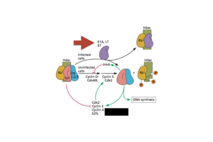
Most cells encountered by viruses are not dividing, and hence do not efficiently support viral DNA synthesis. The genomes of adenoviruses, polyomaviruses, and papillomaviruses encode proteins that cause cells to divide. This effect allows for efficient viral replication, because a dividing cell is producing the machinery for DNA synthesis. Under certain conditions, infections by these viruses do not kill cells, yet they continue to divide due to the presence of viral oncoproteins. Such incessant division gives the cells new properties – they are called transformed cells – and they may eventually become a tumor.

These so-called viral oncoproteins include large T antigen (of SV40, a polyomavirus); E6  and E7 (papillomavirus), and E1A (adenovirus). These viral proteins kick cells into mitosis by inactivating cell proteins (such as Rb, pictured) that are normally involved in regulating cell growth. The cells divide, and in the process produce proteins involved in DNA replication, which are then used for viral replication. These oncoproteins accidentally cause tumors: the replication of none of these viruses is dependent on transformation or tumor formation.

Cells transformed with T, E6/E7, or E1A proteins are commonly used in laboratories because they are immortal. An example is the famous HeLa cell line, transformed by human papillomavirus type 18 (which originally infected Henrietta Lacks and caused the cervical tumor that killed her). Another commonly used transformed cell line is 293 (human embryonic kidney cells transformed by adenovirus E1A). It’s been known for some time that when DNA is introduced into normal (that is, not transformed) cells, they respond with an innate response: interferons are produced. In contrast, when DNA is introduced into the cytoplasm of a transformed cell, there is no interferon response.

To understand why HeLa and HEK 293 cell lines did not respond to cytoplasmic DNA, the authors silenced the viral oncogenes by disrupting them with CRISPR/Cas9. The altered cells produced interferon in response to cytoplasmic DNA. Furthermore, they produced new transformed lines by introducing genes encoding E6, E7, E1A, or T into normal mouse embryonic fibroblasts. These new transformed cells failed to respond to cytoplasmic DNA.

Cytoplasmic DNA is detected in cells by an enzyme called cGAS (cyclic guanosine monophosphate-adenosine monophosphate synthase) together with an adaptor protein known as STING (stimulator of interferon genes). When cytoplasmic DNA is detected by this system, the antiviral interferons are produced. The viral oncoproteins were found to directly bind STING, but not cGAS. A five amino acid sequence within E1A and E7 proteins was identified that is responsible for overcoming the interferon response to cytoplasmic DNA. When this sequence was altered, interaction of the oncoprotein with cGAS was reduced, and antagonism of interferon production in response to cytoplasmic DNA was blocked.

These findings provide a new function for the oncoproteins from three DNA tumor viruses: antagonism of the interferon response to cytoplasmic DNA. Normally DNA is present in the cell nucleus, and when it is detected in the cytoplasm, this is a signal that a virus infection is underway. The cytoplasmic DNA is sensed by the cGAS-STING system, leading to interferon production and elimination of infection. A herpesvirus protein has been identified that binds to STING and blocks interferon responses to cytoplasmic DNA. Clearly antagonism of the cGAS-STING DNA sensing system is of benefit to DNA viruses.

An interesting question is what selection pressure drove the evolution of viral oncogenes. One hypothesis, described above, is that they are needed to induce a cellular environment that supports viral DNA synthesis. The other idea, favored by the authors of this new work, is that oncogenes arose as antagonists of innate immune signaling. But I can’t imagine these DNA viruses without oncogenes, because they would not be able to replicate very efficiently. Could both functions have been simultaneously selected for? Why not – the same five amino acid sequence that binds cGAS also binds cellular proteins (such as Rb), disrupting their function and leading to uncontrolled cell growth!

Link to paper

Source: Virology Blog
Listado de emisiones anteriores

No se encontraron entradas.

Your next migraine might be thanks to your mouth microbes

0

His wife, Lucia Cazares, suffers from bad migraine headaches, especially when she eats certain foods.  Gonzalez had been reading about how bacteria in the gut and mouth affect health, and particularly how certain foods were thought to trigger migraines. As he kept connecting dots, it dawned on him that our symbiotic microbes might play a role in the migraine suffering his wife faced.

“Whenever my wife ate fast food hamburgers, pepperoni pizza or salami—any meat with lots of nitrates—she would get a migraine,” says Gonzalez, now a programmer analyst in Knight’s lab at University of California San Diego School of Medicine.

Nitrates, it turns out, are a common dietary trigger for some of the 38 million Americans who get migraines. Nitrates are also found in cardiovascular medicines, because once they are turned into nitric oxide (NO) in the body, that produces vasodilation. Up to 80% of patients taking nitrates report having severe headaches as a side effect. In addition, nitrates are reduced by bacteria in our mouths into nitrites, which can then be converted to NO by the body. But exactly how nitrates might cause headaches or migraines is unknown.

As Gonzalez and the team were forming the survey for the American Gut Project—a crowd-funded project to sequence microbiomes from citizen scientists—they decided to include questions not only about diet, but also whether participants suffered from migraines.  This would let them look at the oral and gut microbiomes of migraineurs to find if there were significant differences in the bacterial taxa present.

Using 172 oral samples and 1,996 fecal samples from American Gut Project healthy subjects, the team compared sequences from migraineurs and non-migraineurs.  “We didn’t find many differences in the composition of their microbiomes,” says Gonzalez.

So, next, the team decided to look at the likely genes represented by the species that were present to see if there were differences among the genes that code for nitrate-, nitrite- and NO-reducing enzymes. Using the PICRUSt bioinformatic tool, the team found that, particularly in the oral samples, there were significantly more of these genes predicted to be present in migraineurs compared to non-migraineurs.  Although the team did not actually sequence these genes directly from the samples, a statistical test revealed that their predictions were quite robust. Their results are published in mSystems.

“Bacterial genomes have nitrate reductases, enzymes that are the first step to producing the bioactive storage pool of NO in humans,” says Embriette Hyde, project manager for the American Gut Project and co-author on the study. “We already knew that nitrate-reducing bacteria are in the oral cavity and produce measurable effects on blood pressure, and now we have a potential connection to migraines as well.”

Gonzalez and Hyde note that the next step will be to look in patients who suffer from the different type of migraines and see if gene activity correlates to migraine status.  “Then we can start thinking about how to address the problem of migraines,” he says. That might be finding a way to reduce the amount of nitrate-reducers present in a patient’s mouth microbiome. But, it will also be important to find the right balance of microbes to promote cardiovascular health as well.

“My wife and I now joke that we don’t have to worry about her heart, but we do have to worry about her migraines,” says Gonzalez. Perhaps, he says, we will eventually have a “magical probiotic mouthwash” to balance the microbes in our mouths. But, he notes, the real take home message for migraine sufferers is that this is another piece of evidence that they should avoid nitrate triggers in their diet when possible. He advises, “Read food labels and avoid foods with added nitrates.”

Source: ASM Communications

NIH study determines key differences between allergic and non-allergic dust mite proteins

0

This study is the first to provide specific information about the characteristics of dust mite proteins, and may help researchers uncover factors that lead to the development of dust mite allergy and assist in the design of better allergy therapies.

The results, partially funded by the National Institute of Environmental Health Sciences (NIEHS), the National Institute of Allergy and Infectious Diseases, and the National Institute of General Medical Sciences, appeared online Oct. 19 in the Journal of Allergy and Clinical Immunology.

“Allergy researchers have pondered what distinguishes an allergen from a non-allergen for years,” said NIEHS Staff Scientist Geoffrey Mueller, Ph.D., corresponding author of the paper. “There was anecdotal evidence in the field before, and it made sense to examine the organism that causes a lot of allergic sensitization in humans, the common dust mite.”

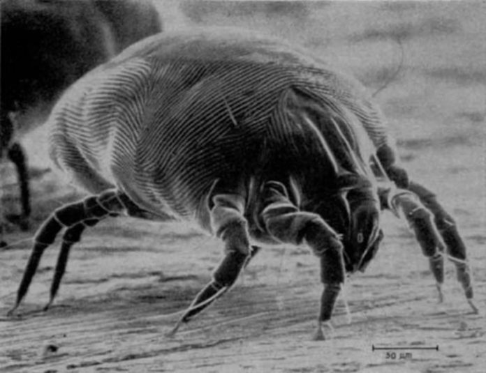
Mueller and his collaborators used two approaches to tackle the question. The first used whole body extracts of dust mites to measure the amount of proteins being made. As a group, the allergens were much more highly produced than non-allergen dust mite proteins.

The second approach involved Duke University scientist Michael Fitzgerald, Ph.D., who utilized a large-scale, mass spectrometry technique to measure the stability of many proteins at once. His team evaluated 656 non-allergens and 19 allergens, and found that the 19 allergens were statistically more stable than dust mite proteins in general.

“I am excited that the analytical methodology we spent so many years developing, turned out to be so useful in addressing this fundamental question about allergenic proteins,” Fitzgerald said.

The finding that dust mite allergens are more durable and more abundant than other dust mite proteins supports two hypotheses about how allergenic compounds stimulate an immune response in the body.

Mueller said one theory is that proteins that are more stable do not break down during the journey from the source, such as dust mite, cockroach, and ragweed pollen, to a person. Another possible explanation is that more stable proteins are harder for the immune system to digest, therefore initiating signals in the body that indicate they are dangerous particles.

Although the usual symptoms of sneezing, red or watery eyes, and nasal congestion, seen in allergies are unwelcome, they are, for the most part, lessened by prescription and over-the-counter allergy medications. However, for the millions of asthmatics who are allergic to dust mites, interacting with dust mite allergens can spur emergency room visits.

“Dust mite allergy is a risk factor for asthma, which is a disease of enormous public health importance in the U.S. and abroad,” said NIEHS Scientific Director Darryl Zeldin, M.D. “Studies such as this one, which enhance our understanding of the characteristics and biology of dust mite allergens, have significant potential to lead to development of new approaches that treat this condition.”

Source: NIH

New Method Detects Multiple Diseases Via DNA Released From Dying Cells Into Blood

0

In a series of experiments involving 320 patients and controls, researchers developed a blood test that can detect multiple pathologies, including diabetes, cancer, traumatic injury and neurodegeneration, in a highly sensitive and specific manner. The novel method infers cell death in specific tissue from the methylation patterns of circulating DNA that is released by dying cells.

The findings are reported in a paper published in the Proceedings of National Academy of Sciences USA, entitled “Identification of tissue specific cell death using methylation patterns of circulating DNA”. The research was performed by an international team led by Dr. Ruth Shemer and Prof. Yuval Dor from The Hebrew University of Jerusalem, and Prof. Benjamin Glaser from Hadassah Medical Center.

Cell death is a central feature of human biology in health and disease. It can signify the early stages of pathology (e.g. a developing tumor or the beginning of an autoimmune or neurodegenerative disease), mark disease progression, reflect the success of therapy (e.g. anti cancer drugs), identify unintended toxic effects of treatment and more. However to date, it is not possible to measure cell death in specific human tissues non-invasively.

The new blood test detects cell death in specific tissues by combining two important biological principles. First, dying cells release fragmented DNA to the circulation, where it travels for a short time. This fact has been known for decades; however since the DNA sequence of all cells in the body is identical, it has not been possible to determine the tissue of origin of circulating DNA, and simple measurements of the amount of circulating DNA is of very limited use. The second principle is that the DNA of each cell type carries a unique chemical modification called methylation. Methylation patterns of DNA account for the identity of cells (the genes that they express), are similar among different cells of the same type and among individuals, and are stable in healthy and disease conditions. For example, the DNA methylation pattern of pancreatic cells differs from the pattern of all other cell types in the body.

The researchers have identified multiple DNA sequences that are methylated in a tissue-specific manner (for example, unmethylated in DNA of neurons and methylated elsewhere), and can serve as biomarkers for the detection of DNA derived from each tissue. They then developed a method to detect these methylated patterns in DNA circulating in blood, and demonstrated its utility for identifying the origins of circulating DNA in different human pathologies, as an indication of cell death in specific tissues. They were able to detect evidence for pancreatic beta-cell death in the blood of patients with new-onset type 1 diabetes, oligodendrocyte death in patients with relapsing multiple sclerosis, brain cell death in patients after traumatic or ischemic brain damage, and exocrine pancreas cell death in patients with pancreatic cancer or pancreatitis.

“Our work demonstrates that the tissue origins of circulating DNA can be measured in humans. This represents a new method for sensitive detection of cell death in specific tissues, and an exciting approach for diagnostic medicine” said Dr. Ruth Shemer of the Hebrew University, a DNA methylation expert and one of the lead authors of the new study.

The approach can be adapted to identify cfDNA derived from any cell type in the body, offering a minimally-invasive window for monitoring and diagnosis of a broad spectrum of human pathologies, as well as better understanding of normal tissue dynamics.

“In the long run, we envision a new type of blood test aimed at the sensitive detection of tissue damage, even without a-priori suspicion of disease in a specific organ. We believe that such a tool will have broad utility in diagnostic medicine and in the study of human biology,” said Prof. Benjamin Glaser, head of Endocrinology at Hadassah Medical Center and another lead author of the study.

The work was performed by Hebrew University students Roni Lehmann-Werman, Daniel Neiman, Hai Zemmour, Joshua Moss and Judith Magenheim, aided by clinicians and scientists from Hadassah Medical Center, Sheba Medical Center and from institutions in Germany, Sweden, the USA and Canada who provided precious blood samples of patients.

Support for the research came from the Juvenile Diabetes Research Foundation, the Human Islet Research Network of the NIH, the Sir Zalman Cowen Universities Fund, the DFG (a Trilateral German-Israel-Palestine program), and the Soyka pancreatic cancer fund.

The Institute for Medical Research-Israel Canada (IMRIC), in the Hebrew University of Jerusalem’s Faculty of Medicine, is one of the most innovative biomedical research organizations in Israel and worldwide. IMRIC brings together the most brilliant scientific minds to find solutions to the world’s most serious medical problems, through a multidisciplinary approach to biomedical research. More information at http://imric.org.

The Hebrew University of Jerusalem is Israel’s leading academic and research institution, producing one-third of all civilian research in Israel. For more information, visit http://new.huji.ac.il/en.

Hadassah-Hebrew University Medical Center is Israel’s leading academic hospital, combining the highest quality of medical care with world-class basic and translational research.  For more information, visit http://www.hadassah-med.com.

Source: new.huji.ac.il

A bacterial role in breast cancer development and prevention

0

Microbial infection is implicated in an ever-growing number of types of cancer. Adding to the already long list of microbial-associated cancers, an increasing body of evidence suggests breast cancer may also be associated with a specific microbial milieu. A report in Applied and Environmental Microbiology confirms that the breast tissue microbial inhabitants differ between women with and without breast cancer.

Could bacterial colonization be one of the many factors that increase risk of developing breast cancer? To test the microbiome of different types of breast tissue, first author Camilla Urbaniak and senior scientist Gregor Reid received samples from 71 women: 45 with cancerous tumors, 13 with noncancerous tumors, and 23 with no tumors. The scientists examined the breast tissue surrounding the tumors, and not the tumors themselves. The bacteria associated with the tissues were assessed by 16S rRNA gene sequencing after amplification.

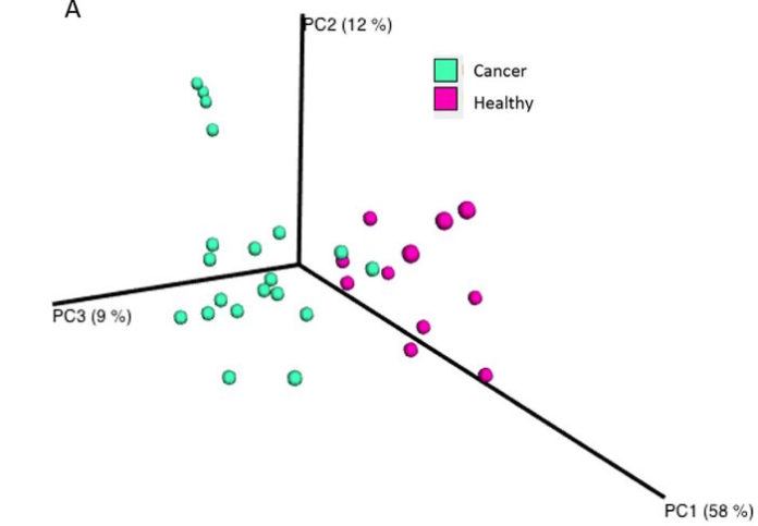
The scientific team observed a clear difference in microbial inhabitants of the women with breast cancer compared to the women without. Using a UniFrac plot (see right), the researchers measured the similarity between samples and plotted it as a measure of distance from other samples. The samples from the women with cancer (green circles) and from the women without cancer (red circles) form separate clusters, showing a similarity within the group and differences from the other group.

What bacteria did they find? Among other differences, the scientists observed that women with cancer showed higher levels of Escherichia coli and Staphylococcus aureus, while the women without cancer showed higher levels of Lactobacillus and Streptococcus species.

Some E. coli strains harbor a pks pathogenicity island in their genome, and pks-containing E. coli have been implicated with some colon cancers due to their ability to cause double-stranded DNA breaks in surrounding host cells. An increased number of DNA breaks also increases opportunities for a poorly repaired break, which may add to cancerous cell proclivities (since cancer is often a disease of multiple mutations). Using the pks E. coli isolated from the breast tissue, the research team confirmed that this strain also increases double-stranded DNA breaks in cell culture.

Can women influence their breast microbiome to prevent high levels of E. coli or S. aureus colonization? Studies support that drinking microbially fermented products, such as kefir, is associated with lower risk of breast cancer. Orally ingested Lactobacillus bacteria protect animals from experimentally developing cancer ($), but there are no results supporting this claim yet in humans. Future studies may demonstrate a role for probiotics as a preventative measure against breast cancer.

Skeptical readers may be hesitant to conclude that breast tissue bacteria contribute to cancer, and there are many additional experiments to show causation still needed. However, if the link is an association caused by the cancer itself, the findings still provide a noninvasive way to monitor women for their breast cancer risk that might assist breast examinations and mammograms, as pictured at the top (source) as screens for early cancerous growth. For younger women (those under 45), mammograms are recommended after discussing other risk factors, such as family history of cancer. Characterizing one’s breast microbiome may inform the decision to schedule a mammogram.

It took Reid and his colleagues years to convince the scientific world that breast tissue is a niche that harbors microbes. This research confirms his team’s earlier work and identifies bacteria that may play a role in breast cancer development. The hope is that these discoveries will move into therapeutics, diagnostics, and preventative measures to protect the 1 in 8 women who will develop breast cancer during their lifetimes.

Source: ASM

High-protein diet curbs metabolic benefits of weight loss

0

But in a study of 34 postmenopausal women with obesity, researchers at Washington University School of Medicine in St. Louis found that eating too much protein eliminates an important health benefit of weight loss: improvement in insulin sensitivity, which is critical to lowering diabetes risk.

The findings are available Oct. 11 in the journal Cell Reports.

“We found that women who lost weight eating a high-protein diet didn’t experience any improvements in insulin sensitivity,” said principal investigator Bettina Mittendorfer, professor of medicine. “However, women who lost weight while eating less protein were significantly more sensitive to insulin at the conclusion of the study. That’s important because in many overweight and obese people, insulin does not effectively control blood-sugar levels, and eventually the result is type 2 diabetes.”

Insulin sensitivity is a good marker of metabolic health, one that typically improves with weight loss. In fact, the women in the study who lost weight while consuming less protein experienced a 25 to 30 percent improvement in their sensitivity to insulin.

Mittendorfer and her colleagues studied 34 women with obesity who were 50 to 65 years of age. Although all of the women had body mass indices (BMI) of at least 30 — a BMI of 30 or more indicates significant obesity — none had diabetes.

The participants were randomly placed into one of three groups for the 28-week study. In the control group, women were asked to maintain their weight. In another group, the women ate a weight-loss diet that included the recommended daily allowance (RDA) of protein: 0.8 grams per kilogram of body weight. For a 55-year-old woman who weighs 180 pounds, that would come to about 65 grams of protein per day.

In the third group, the women ate a diet designed to help lose weight, but they consumed more protein, taking in 1.2 grams per kilogram of body weight, or almost 100 grams for that same 180-pound woman.

“We provided all of the meals, and all the women ate the same base diet,” Mittendorfer explained. “The only thing we modified was protein content, with very minimal changes in the amount of fat or carbohydrates. We wanted to hone in on the effects of protein in weight loss.”

The researchers focused on protein because in postmenopausal women, there is a common belief that consuming extra protein can help preserve lean tissue, keeping them from losing too much muscle while they lose fat.

“When you lose weight, about two-thirds of it tends to be fat tissue, and the other third is lean tissue,” Mittendorfer said. “The women who ate more protein did tend to lose a little bit less lean tissue, but the total difference was only about a pound. We question whether there’s a significant clinical benefit to such a small difference.”

The women who ate the recommended amount of protein saw big benefits in metabolism, led by a 25 to 30 percent improvement in their insulin sensitivity. Such improvements lower the risk for diabetes and cardiovascular disease. The women on the high-protein diet, meanwhile, did not experience those improvements.

“Changing the protein content has very big effects,” Mittendorfer said. “It’s not that the metabolic benefits of weight loss were diminished — they were completely abolished in women who consumed high-protein diets, even though they lost the same, substantial amounts of weight as women who ate the diet that was lower in protein.”

It’s still not clear why insulin sensitivity didn’t improve in the high-protein group, and Mittendorfer said it’s not known whether the same results would occur in men or in women already diagnosed with type 2 diabetes. She plans to continue researching the subject.

Source: Science Blog

Circadian rhythm-related genes: implication in autoimmunity and type 1 diabetes

0

There is growing evidence about the implication of the circadian rhythm in diabetes development. Studies in mice have shown that the disruption of circadian rhythms can accelerate diabetes and -cell loss.

In humans a link between the central circadian rhythm regulation and glucose homeostasis has been suggested by findings such as the polymorphism in MTNR1B, encoding the melatonin receptor 1B, that increases the risk for type 2 diabetes. Transcription and translation of core clock components circadian locomotor output cycles kaput (CLOCK), aryl hydrocarbon receptor nuclear translocator-like 1 (ARNTL1), aryl hydrocarbon receptor nuclear translocator-like 2 (ARNTL2), period circadian proteins (PER1, PER2, PER3) and Cryptochromes (CRY1 and CRY2) play a pivotal role in rhythm generation in the suprachiasmatic nucleus, which is the site of the master circadian oscillator in mammals, but also in the control of peripheral oscillations.

Authors: B. Lebailly1,2, C. Boitard1 & U. C. Rogner1

  1. Institut Cochin (INSERM U1016, CNRS UMR-S8104, Département “Endocrinologie, Métabolisme et Diabètes), Paris, France
  2. Cellule Pasteur, University Pierre and Marie Curie, Paris, France

Read the full text here.

El Microscopio – Program 25

0
  • Dr. Elena Verdú (Canada). Interview about the role of host-microbial and celiac disease.
  • Dr. Jason Park (USA). Interview about cancer genomics.
  • Dr. Karl Bacos (Sweden). Interview about a new method that measures the risk of type 2 diabetes in blood
  • Agenda.
  • News and events about clinical chemistry.

Interview: Dr. Jason Park (USA)

0

Jason Park, MD, PhD, FCAP is an Associate Professor in the Department of Pathology and the Eugene McDermott Center for Human Growth and Development at UT Southwestern Medical School. He is the Medical Director of the Advanced Diagnostics Laboratory at Children’s Health, Children’s Medical Center Dallas. Dr. Park’s clinical and research interests are in genomic medicine, gastrointestinal diseases, and clinical informatics. He is the author/co-author of over 100 journal articles, book chapters and abstracts. He serves as a member of the Editorial Boards of Clinical Chemistryand Archives of Pathology & Laboratory Medicine. His research has resulted in 13 issued U.S. patents on cancer diagnostics, cancer therapeutics and nanotechnology.

Dr. Park trained in a combined MD and PhD program at Thomas Jefferson University and was a pathology resident and chief resident in the Department of Pathology and Laboratory Medicine at the Hospital of the University of Pennsylvania. After residency, he was a clinical fellow in gastrointestinal and liver pathology at the Johns Hopkins Hospital. He is certified by the American Board of Pathology (clinical informatics, clinical pathology and anatomic pathology) as well as the American Board of Clinical Chemistry (molecular diagnostics).

Agenda


Radio El Microscopio

Ze Xiong

Últimas notas publicadas