Inicio Blog Página 20

UNC Researchers identify a new HIV reservoir

0
J. Victor Garcia, PhD, professor of medicine, microbiology and immunology at UNC School of Medicine

Macrophages are large white blood cells found in tissues throughout the body including the liver, lung, bone marrow and brain. The discovery of this additional viral reservoir has significant implications for HIV cure research. These findings were published in Nature Medicine on Monday, April 17.

“These results are paradigm-changing because they demonstrate that cells other than T cells can serve as a reservoir for HIV,” said Jenna Honeycutt, PhD, lead author and postdoctoral research associate in the UNC division of infectious diseases. “The fact that HIV-infected macrophages can persist means that any possible therapeutic intervention to eradicate HIV might have to target two very different types of cells.”

Last spring, this laboratory lead by J. Victor Garcia, PhD, professor of medicine, microbiology and immunology at UNC School of Medicine, demonstrated the ability of tissue macrophages to support HIV replication in vivo in the total absence of human T cells. But how macrophages would respond to antiretroviral therapy (ART) and whether macrophages represented a reservoir for HIV after treatment were unknown.

Macrophages are myeloid lineage cells that have been implicated in HIV pathogenesis and in the trafficking of virus into the brain. Using a humanized myeloid-only mouse (MoM) model devoid of T cells, Garcia and his team showed that ART strongly suppresses HIV replication in tissue macrophages. Yet when HIV treatment was interrupted, viral rebound was observed in one third of the animals. This is consistent with the establishment of persistent infection in tissue macrophages.

“This is the first report demonstrating that tissue macrophages can be infected and that they respond to antiretroviral therapy,” Honeycutt said. “In addition, we show that productively infected macrophages can persist despite ART; and most importantly, that they can reinitiate and sustain infection upon therapy interruption even in the absence of T cells – the major target of HIV infection.”

Now that Garcia and his team know HIV persists in macrophages, the next steps will be to determine what regulates HIV persistence in tissue macrophages, where in the body persistently infected macrophages reside during HIV treatment and how macrophages respond to possible therapeutic interventions aimed at eradicating HIV from the body.

The UNC School of Medicine team collaborated with scientists in UNC’s department of biostatistics, the theoretical division at Los Alamos National Laboratory, Veterans Affairs San Diego Healthcare System, and the departments of medicine and pathology at the University of California at San Diego. This study was funded by the National Institute of Mental Health and the National Institute of Allergy and Infectious Diseases of the U.S. National Institutes of Health.

Jenna Honeycutt, PhD, lead author and postdoctoral research associate in the UNC division of infectious diseases

The mission of UNC’s Institute for Global Health & Infectious Diseases is to harness the full resources of the University and its partners to solve global health problems, reduce the burden of disease, and cultivate the next generation of global health leaders. Learn more at www.globalhealth.unc.edu.

Source: unchealthcare.org
Listado de emisiones anteriores

No se encontraron entradas.

European Federation of Laboratory Medicine e-learning platform

0

European Federation of Laboratory Medicine e-learning platform offers:

  • webinars
  • recordings of presentations (from webinars and congresses) packed with didactic quizzes into an e-learning courses
  • e-learning courses (educational activities of EFLM WG´s, postgradual education of trainees  – Specialists in Laboratory Medicine …)

List of EFLM webinars (e-seminars)

  • November 14, 2017. Harmonization of preanalytical phase in Europe. Ana-Maria Simundic
  • October 19, 2017. Hepatic fibrosis assessment using multiparametric biomarker tests. Ralf Lichtinghagen
  • September 26, 2017. EFLM webinar – Reliable estimates of biological variation – the way forward. Aasne Karine Aarsand
  • December 13, 2016. EFLM webinar – Patient with shock and multiorgan failure. Anna Merino
  • November 17, 2016. EFLM webinar – Biomarkers in guiding treatment of heart failure. Alan S. Maisel
  • June 7, 2016. EFLM webinar – Non-fasting lipid profiles: implications for lipoprotein measurement and reporting. Michel Langlois
  • May 10, 2016. EFLM webinar – The estimate of measurement uncertainty. Ilenia Infusino
  • April 26, 2016. EFLM webinar – Rational use of laboratory tests. Gustav Kovac

Web Page: elearning.eflm.eu

FOCiS 2017

0

FOCIS is the meeting in translational immunology that will give you a competitive edge in your career. Stay ahead of the curve with leading clinicians and researchers delivering the latest breakthroughs across immune-mediated diseases. Focusing on molecular pathways and their implications in human disease provides the unique opportunity for innovative thinking and apply ideas from the pathologies of other diseases to uncover novel solutions to challenges in the diseases you study.

Meeting Venue: Hilton Chicago, 720 South Michigan Avenue, Chicago, IL 60605. (312) 9224400

Continuing Medical Education (CME) In the past, FOCIS has made it possible for meeting participants to obtain CME credit for attending the annual meeting. In order to enable all those involved in the research and treatment of immunemediated diseases to participate in the exchange of ideas that contribute to the forward motion of the field, FOCIS will not offer CME credit for FOCIS 2017. Certificates of attendance will be available upon request.

Full Schedule

Visit de FOCIS 2017 microsite to register, book a room and more!: eventscribe.com/2017/FOCIS

Biomarkers and Immuno-Oncology World Congress

0

Cambridge Healthtech Institute’s Thirteenth Annual Biomarkers and Immuno-Oncology World Congress is the longest-running event dedicated to all areas of biomarker research and development spanning the pharmaceutical and diagnostic pipelines. With the promising developments in immuno-oncology over the past five years, much of the biomarker R&D has shifted to discovery and development of predictive biomarkers for immunotherapy patient stratification, understanding tumor mutational load and neoantigens in developing personalized immunotherapy approaches, using biomarker or mechanism data to choose rational combination immunotherapy, and applying immune profiling to predict response to immunotherapy.

The Biomarkers and Immuno-Oncology World Congress brings together a unique and international mix of large and medium pharmaceutical, biotech and diagnostics companies, leading universities and clinical research institutions, government and national labs, CROs, emerging companies and tool providers—making the Congress a perfect meeting-place to share experience, foster collaborations across industry and academia, and evaluate emerging technologies. Now in its thirteenth year, the Biomarkers and Immuno-Oncology World Congress is the leading annual meeting dedicated to biomarkers, diagnostics, and immuno-oncology research and implementation that consistently delivers a cutting-edge agenda, 400+ senior delegates, and a sold-out exhibit hall.

SHORT COURSES*

  • MONDAY AFTERNOON, MAY 1 | 1:00 – 4:00 PM
    • SC1: FIT-FOR-PURPOSE BIOMARKER ASSAY DEVELOPMENT AND VALIDATION
  • MONDAY EVENING, MAY 1 | 5:00 – 8:00 PM
    • Dinner Workshop. SC2: LIQUID BIOPSY FOR IMMUNO-ONCOLOGY AND PRECISION MEDICINE
    • Dinner Short Course. SC3: PREPARING FOR COMPANION DIAGNOSTIC DEVICE STUDIES AND SUBMISSIONS TO FDA
  • TUESDAY EVENING, MAY 2 | 6:00 – 9:00 PM
    • Dinner Short Course. SC4: NEXT-GENERATION SEQUENCING AS A CLINICAL TEST
    • Dinner ThinkTank. SC5: PD-L1 ASSAYS FOR BIOMARKERS AND COMPANION DIAGNOSTICS
  • WEDNESDAY EVENING, MAY 3 | 6:15 – 9:15 PM
    • Dinner Executive ThinkTank. SC6: COMPLEMENTARY DIAGNOSTICS
    • Dinner Short Course. SC7: IMMUNE MONITORING IN CANCER

More Information: biomarkerworldcongress.com

Lymphatic filariasis

0

The disease

Lymphatic filariasis, commonly known as elephantiasis, is a neglected tropical disease. Infection occurs when filarial parasites are transmitted to humans through mosquitoes. Infection is usually acquired in childhood causing hidden damage to the lymphatic system.

The painful and profoundly disfiguring visible manifestations of the disease, lymphoedema, elephantiasis and scrotal swelling occur later in life and can lead to permanent disability. These patients are not only physically disabled, but suffer mental, social and financial losses contributing to stigma and poverty.

Currently, 947 million people in 54 countries are living in areas that require preventive chemotherapy to stop the spread of infection.

The global baseline estimate of persons affected by lymphatic filariasis was 25 million men with hydrocele and over 15 million people with lymphoedema. At least 36 million persons remain with these chronic disease manifestations. Eliminating lymphatic filariasis can prevent unnecessary suffering and contribute to the reduction of poverty.

Cause and transmission

Lymphatic filariasis is caused by infection with parasites classified as nematodes (roundworms) of the family Filariodidea. There are 3 types of these thread-like filarial worms:

  • Wuchereria bancrofti, which is responsible for 90% of the cases
  • Brugia malayi, which causes most of the remainder of the cases
  • Brugia timori, which also causes the disease.

Adult worms lodge in the lymphatic vessels and disrupt the normal function of the lymphatic system. The worms can live for an average of 6–8 years and, during their life time, produce millions of microfilariae (immature larvae) that circulate in the blood.

Mosquitoes are infected with microfilariae by ingesting blood when biting an infected host. Microfilariae mature into infective larvae within the mosquito. When infected mosquitoes bite people, mature parasite larvae are deposited on the skin from where they can enter the body. The larvae then migrate to the lymphatic vessels where they develop into adult worms, thus continuing a cycle of transmission.

Lymphatic filariasis is transmitted by different types of mosquitoes for example by the Culex mosquito, widespread across urban and semi-urban areas, Anopheles, mainly found in rural areas, and Aedes, mainly in endemic islands in the Pacific.

Symptoms

Lymphatic filariasis infection involves asymptomatic, acute, and chronic conditions. The majority of infections are asymptomatic, showing no external signs of infection. These asymptomatic infections still cause damage to the lymphatic system and the kidneys, and alter the body’s immune system.

When lymphatic filariasis develops into chronic conditions it leads to lymphoedema (tissue swelling) or elephantiasis (skin/tissue thickening) of limbs and hydrocele (scrotal swelling). Involvement of breasts and genital organs is common. Such body deformities often lead to social stigma and sub-optimal mental health, loss of income-earning opportunities and increased medical expenses for patients and their caretakers. The socioeconomic burdens of isolation and poverty are immense.

Acute episodes of local inflammation involving skin, lymph nodes and lymphatic vessels often accompany chronic lymphoedema or elephantiasis. Some of these episodes are caused by the body’s immune response to the parasite. Most are the result of secondary bacterial skin infection where normal defences have been partially lost due to underlying lymphatic damage. These acute attacks are debilitating, may last for weeks, and are the primary cause of lost wages among persons suffering with lymphatic filariasis.

Source: WHO

For immune cells, discrimination is a good thing

0

The training process teaches immune cells to discriminate between tissues of the human host – “self” cells – and proteins from a deleterious pathogen – “non-self.” Knowing the difference between the two of these entities prevents friendly fire but also ensures the immune system is prepared to launch an attack against a pathogen if necessary.

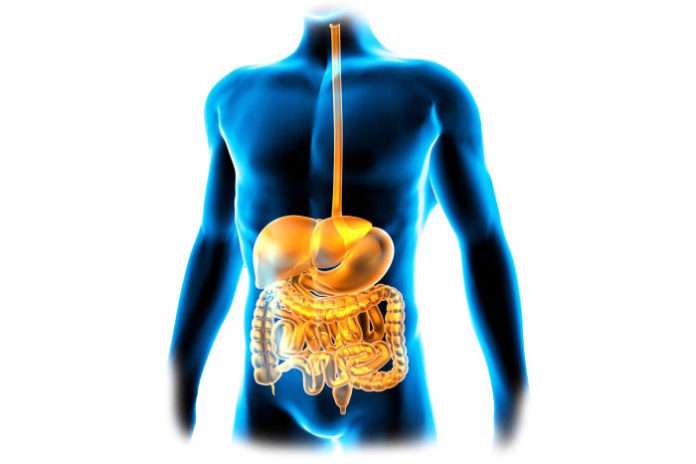
Despite receiving ample training, it is more difficult for immune cells in the epithelial lining of the gastrointestinal (GI) tract to make the distinction between self and non-self than it is for others because of their regular interactions with the microbiome, the “good” bacteria. Gut bacteria won’t be registered by the immune cells as “self,” but they still cannot be attacked without serious health repercussions.

The population of bacteria in the GI tract, called the “gut microbiome,” are part of a large community of beneficial bacteria in the body that influence health in numerous ways. A recent discovery from the new LMU study identified a mechanism that helps immune cells maintain a sort of “immunological equilibrium” in the GI tract, both protecting the good bacteria and defending against the bad bacteria.

The new mechanism clearly separates two opposite roles of immune players called dendritic cells (DCs). On one hand, the immune system counts on DCs to launch an immune attack in response to a pathogenic infection. On the other hand, DCs are also charged with promoting immunological tolerance, suppressing the immune response when needed.

DCs suppress the immune system by stimulating regulatory T cells (Tregs) that, as their name suggests, regulate the tolerance process and inhibit various inflammatory parts of the immune system. In the GI tract, epithelial DCs “recognize and internalize microbial proteins” before they travel to intestinal lymph nodes.

Once in the nodes, bacterial proteins are processed into small fragments which are displayed on the surface of the DCs as a way to flag down Tregs. Then, Tregs recognize the surface markers as a signal to begin suppressing any immune response against the specific markers held on the surface of the DCs, a reaction researchers believe to be unique to the particular marker being held on the cell surface.

This interactive behavior between DCs, Tregs, and microbial proteins from the GI tract is continuous, with Tregs and the immune system receiving a constant update of the goings-on in the gut microbiome, but a disruption can cause DCs to switch from suppressive behavior to stimulative behavior in terms of the immune response.

CD40, a signaling molecule and a surface receptor on DCs, sometimes acts as an alarm. When effector T cells bind CD40 on the surface of DCs, they transition into an immune-stimulatory state instead of the immune-suppressing state associated with Tregs.

LMU researchers demonstrated in mice models the DC transition, which depends on interactions between other immune cells and its surface markers. For example, when researchers triggered permanent CD40 signaling, severe colitis resulted. Colitis is an infection the GI tract characterized by inflammation.

In the case of CD40 signaling, DCs still make the trip from the GI tract epithelial tissue to the lymph nodes, but once they reach the nodes, they die before they can interact with Tregs, failing to initiate suppression of the immune system, resulting in colitis

“These findings show that interaction between dendritic cells and regulatory T-cells is essential for the maintenance of the correct immunological equilibrium or homeostasis in the gut,” explained study leader Professor Thomas Brocker. The study was recently published in the journal Nature Communications.

Source: LabRoots

New approach makes cells resistant to HIV

0

“This protection would be long term,” said Jia Xie, senior staff scientist at TSRI and first author of the study published today in the journal Proceedings of the National Academy of Sciences.

The researchers, led by study senior author Richard Lerner, M.D., Lita Annenberg Hazen Professor of Immunochemistry at TSRI, plan to collaborate with investigators at City of Hope’s Center for Gene Therapy to evaluate this new therapy in efficacy and safety tests, as required by federal regulations, prior to testing in patients.

“City of Hope currently has active clinical trials of gene therapy for AIDS using blood stem cell transplantation, and this experience will be applied to the task of bringing this discovery to the clinic,” said John A. Zaia, M.D., director of the Center for Gene Therapy in the Hematological Malignancy and Stem Cell Transplantation Institute at City of Hope. “The ultimate goal will be the control of HIV in patients with AIDS without the need for other medications.”

“We at TSRI are honored to be able to collaborate with physicians and scientists at City of Hope, whose expertise in transplantation in HIV patients should hopefully allow this therapy to be used in people,” added Lerner.

The new TSRI technique offers a significant advantage over therapies where antibodies float freely in the bloodstream at a relatively low concentration. Instead, antibodies in the new study hang on to a cell’s surface, blocking HIV from accessing a crucial cell receptor and spreading infection.

Xie called it the “neighbor effect.” An antibody stuck nearby is more effective than having many antibodies floating throughout the bloodstream. “You don’t need to have so many molecules on one cell to be effective,” he said.

Before testing their system against HIV, the scientists used rhinovirus (responsible for many cases of the common cold) as a model. They used a vector called lentivirus to deliver a new gene to cultured human cells. This gene instructed cells to synthesize antibodies that bind with the human cell receptor (ICAM-1) that rhinovirus needs. With the antibodies monopolizing that site, the virus cannot enter the cell to spread infection.

“This is really a form of cellular vaccination,” said Lerner. Because the delivery system can’t reach exactly 100 percent of cells, the finished product was a mix of engineered and unengineered cells. The researchers then added rhinovirus to these cell populations and waited to see what would happen.

The vast majority of cells died in about two days. In dishes with only unengineered cells, the population never recovered. There was an initial die-off in the mixed engineered/unengineered populations, too, but their numbers quickly bounced back. After 125 hours, these cell populations were back up to around the same levels as cells in an undiseased control group.

In essence, the researchers had forced the cells to compete in Darwinian, “survival-of-the-fittest” selection in a lab dish. Cells without antibody protection died off, leaving protected cells to survive and multiply, passing on the protective gene to new cells.

This success led the researchers to test the same technique against HIV. To infect a person, all strains of HIV need to bind with a cell surface receptor called CD4. So the scientists tested antibodies that could potentially protect this receptor on the very immune cells normally killed by HIV. “This research is possible because of the ability to select specialized antibodies from combinatorial antibody libraries,” said Lerner.

Again, their technique worked. After introducing cells to the virus, the researchers ended up with an HIV-resistant population. The antibodies recognized the CD4 binding site, blocking HIV from getting to the receptor.

The scientists further confirmed that these tethered antibodies blocked HIV more effectively than free-floating, soluble antibodies in experiments led by study co-authors Devin Sok of the International AIDS Vaccine Initiative (IAVI) and TSRI Professor Dennis R. Burton, who is also scientific director of the IAVI Neutralizing Antibody Center and of the National Institutes of Health’s Center for HIV/AIDS Vaccine Immunology and Immunogen Discovery (CHAVI-ID) at TSRI.

Joseph Alvarnas, M.D., director of Value-Based Analytics at City of Hope, explained how the TSRI technique could help patients, who—despite treatment with anti-retroviral drugs—still suffer from higher rates of diseases, such as cancers. “HIV is treatable but not curable—this remains a disease that causes a lot of suffering. That makes the case for why these technologies are so important,” he said.

In addition to potentially collaborating with City of Hope, Xie said the next step in this research is to try engineering to protect a different receptor on the cell surface.

More information: Immunochemical engineering of cell surfaces to generate virus resistance, Proceedings of the National Academy of Sciences (2017). www.pnas.org/cgi/doi/10.1073/pnas.1702764114

Source: medicalxpress.com

Could celiac disease be triggered by a common virus?

0

Researchers have demonstrated how a seemingly benign human virus can trigger an immune response against gluten when the protein is given to mice. Their study is published in the journal Science.

They found that intestinal viruses can cause the immune system to overreact to gluten, and wondered if this could be a trigger to developing celiac disease. They tested this by infecting mice with a common strain of reovirus, which is usually symptomless, and then feeding them small amounts of gliadin, a component of gluten.

Their results showed that the mice infected with the virus produced up to three times more antibodies against the protein in the two days after they consumed it than when they were virus-free.

The researchers also looked at patients with celiacs disease and found they had much higher levels of antibodies against reoviruses than those without the disease. Those with higher levels of antibodies also had higher levels of the molecule IRF-1, which plays a key role in the loss of gluten tolerance.

The study concludes that infection from a reovirus could be key in developing celiac disease, especially among babies. The researchers suggest that when children first encounter solid food, usually around six months, they are particularly vulnerable to viral infections because their immune system is still developing.

For those babies already genetically predisposed to celiac disease, the combination of being vulnerable to viruses coupled with their first exposure to gluten might cause a perfect storm to develop the disease. “That’s why we believe that once we have more studies, we may want to think about whether children at high risk of developing celiac disease should be vaccinated,” said senior author Bana Jabri in a statement.

This new evidence doesn’t mean that every person who insists they need to be gluten-free are now validated. Celiac disease is rare, and even fewer people are diagnosed with it. However, low diagnosis doesn’t necessarily mean people don’t have celiac disease, it just means they haven’t officially been diagnosed with it by a doctor.

With the recent trend for gluten-free food as a “healthy” option, despite there being no evidence that it is, more people are cutting out gluten by self-diagnosis. If you think you might be intolerant to gluten, go and see a doctor first. They can’t diagnose your body’s relationship with gluten if you have already removed it from your diet.

Source: IFLScience

Educational Webinar: Mass Spectrometry in the Clinical Laboratory

0

DATE: April 27, 2017

TIME: 10:00am PT, 1:00pm ET

Presented by: William Clarke, PhD. Associate Professor of Pathology. Johns Hopkins University School of Medicine

Mass spectrometry is increasingly being used for clinical testing and evaluation of patients in various settings. The utilization of this technology is driven by its potential to perform challenging analyses in complex matrices and sample types. While mass spectrometry is a powerful tool, there are also some challenges that must be addressed for it to be routinely used for medical testing. This presentation will discuss the increasing use of mass spectrometry for clinical testing, discuss challenges for implementation of the technology, and present some cases where it may have significant impact in medical care.

Learning Objectives:

  • Describe how mass spectrometry is currently used in clinical laboratories
  • Discuss the challenges regarding implementation of mass spectrometry into routine testing
  • Define areas where mass spectrometry can be used to positively impact medical care

Registration: labroots.com//mass-spectrometry-clinical-laboratory

Agenda


Radio El Microscopio

Ze Xiong

Últimas notas publicadas